Journal of Vita Columbia Volume 2 Issue 1 – Clinical Nutrition

Vitamin B6: A Synopsis of this Critical Micronutrient

January 18th, 2022
How to cite this article:

Latcu CM, Baila BM, Grewal VP. Vitamin B6: A Synopsis of this Critical Micronutrient. 2022. Journal of Vita Columbia. 2(1).

Abstract:

Vitamin B6, first discovered as a factor that cured dermatitis in rats, is a water-soluble vitamin that works as a coenzyme for more than 100 essential enzyme-catalyzed reactions. It is rapidly metabolized and excreted. Therefore it is very unlikely that vitamin B6 deficiency or toxicity happens. In case of toxicity nerve damage-related symptoms and other symptoms can occur. Its most biologically active form, PLP – pyridoxal 5’-phosphate form, and its dependent enzymes play an important role in cellular metabolism and other important reactions. Finally, vitamin B6 is catabolized through the oxidation of pyridoxal to 4-pyridoxic acid, which is excreted in the urine. Besides all these general characteristics of this vitamin, studies also outline new insights into its impact on human health and the use of vitamin B6 supplements.

Introduction:

Vitamin B6 (Vit. B6), also called pyridoxine, part of the group of vitamin B complex, water-soluble, chemically quite distinct compounds, is a really important compound for overall cellular metabolism being involved in amino acid biosynthesis and degradation, also in glucose and fatty acid metabolism. [1]

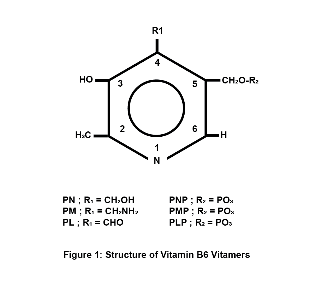
Vit. B6 consists of a group of six related vitamers: pyridoxal (PL), pyridoxine (PN), pyridoxamine (PM) and the 5′-phosphates (PLP- pyridoxal 5’-phosphate, PNP- pyridoxine 5’-phosphate and PMP – pyridoxamine-5’-phosphate). The animal tissues contain mainly the PLP and PMP forms, and plant-based products the PN and PNP forms. Humans have 4-pyridoxic acid (4-PA) as their main excretory form. PN, PL and PM forms are mainly converted to PLP via some consecutive reactions that require a kinase (whose role is to phosphorylate the 5′hydroxymethyl group) and the pyridoxamine phosphate oxidase – PNPO (whose role is to phosphorylate the PNP and the PMP). The PLP is the biologically active form of vitamin B6.  [2][4]

Metabolism of Vitamin B6

Absorption & Transportation

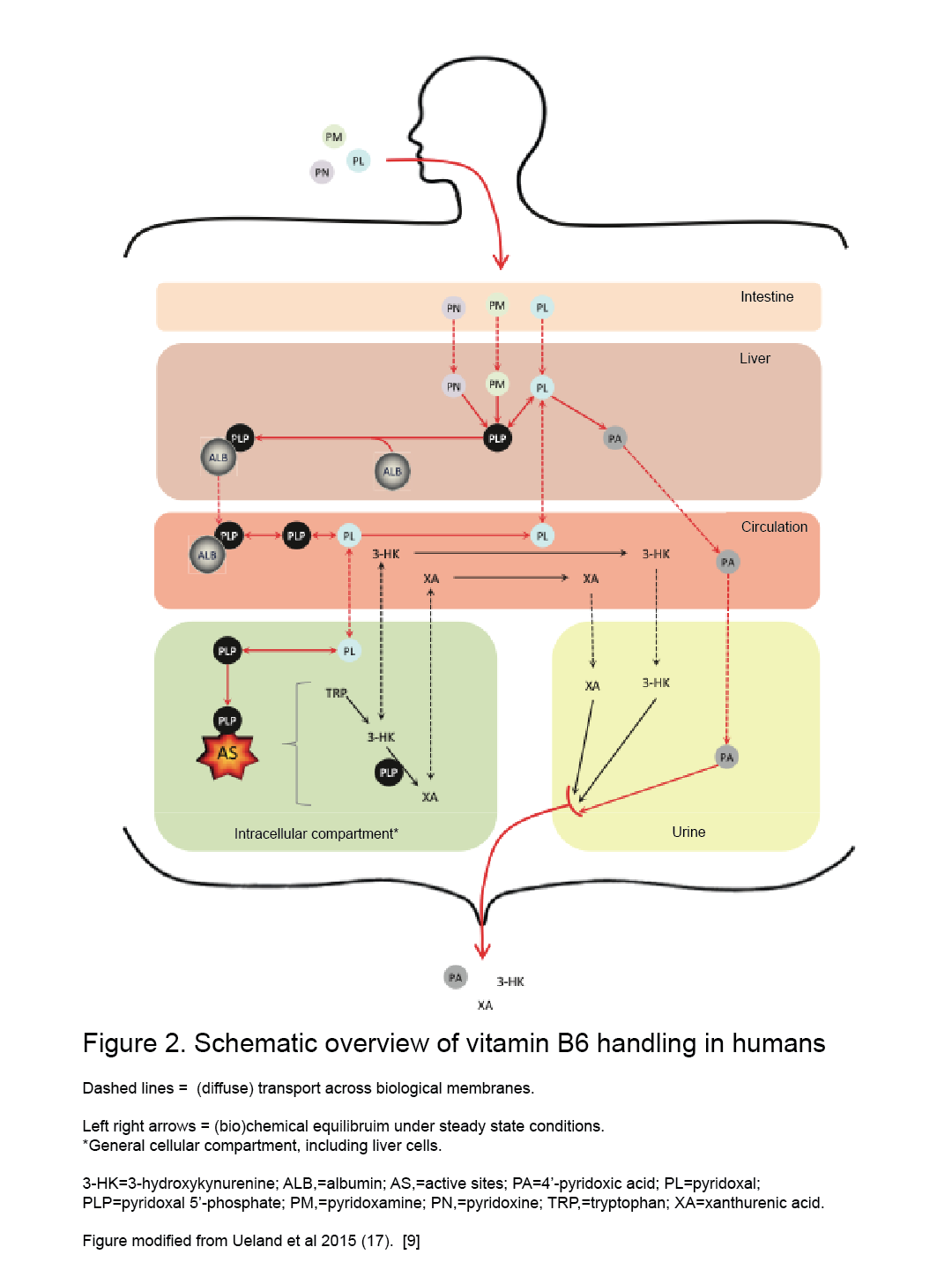
Humans and animals cannot synthesize vit. B6, hence they rely on external sources. [2] The absorption, through passive diffusion, takes place in the intestine and is divided into two parts: dietary ingestion that takes place in the small intestine (jejunum) and the uptake of bacteria produced vit. B6 that takes place in the large intestine. [3][9] The intestine absorbs only the nonphosphorylated B6 vitamers and, in the intracellular compartment of the intestine, most of them go to the liver and are converted to phosphorylated forms (PL to PLP by PL kinase for instance). [2][7][9] Absorption also requires phosphatase-mediated hydrolysis followed by transport of the nonphosphorylated form into the mucosal cell, regardless of dose. [2] PLP must be bound to serum albumin (ALB) as a Schiff base form and be delivered entirely from the liver as a PLP-albumin complex, which protects PLP from hydrolysis. [2][7][8][9] After these steps, the PLP-albumin complex requires dephosphorylation (by an alkaline phosphatase) to pyridoxal (less hydrophilic form) before being secreted into the circulatory blood system for delivery to the different tissues and organs, and even crossing the blood-brain barrier (BBB). [7][8] After the liver, the choroid plexus seems to be the only organ able to readily release PLP, which explains the relatively high percentage of PLP in cerebrospinal fluid. [7] After all these reactions, the B6 vitamers must be rephosphorylated by pyridoxal kinase in the brains cells and in other target cells too, found mostly in the mitochondria and the cytosol (where quite similar mechanisms occur). [2][7] 

There is also a „salvage pathway” [7] for PLP formation from other forms such as PNP and PMP by oxidation by PNP oxidase. [2] PMP form is also generated from PLP via aminotransferase reactions. [2] PLP capacity of binding limits the accumulation in tissues and organs at high intakes of vit. B6. [2] If this capacity is exceeded, free PLP is rapidly hydrolyzed and nonphosphorylated forms of vit. B6 are released by the liver and other tissues into circulation. [2] At pharmacological doses of B6, the high capacities for PLP-protein binding of muscle, plasma, and erythrocytes (hemoglobin) allow them to accumulate very high levels of PLP when other tissues are saturated.[2]

Excretion

B6 vitamers are excreted mainly in the urine, but also in feces. [2][7][9] When the intakes exceed the requirements, dephosphorylation of the pyridoxal phosphate takes part (mainly in the liver) and this is oxidized to the bioactive catabolite 4- pyridoxic acid (PA) – the main excretory product – and other forms or could also be excreted unchanged in urine [2][7][9]

Functions of Vitamin B6:

Based on several studies vit. B6, in the form of pyridoxal 5’-phosphate (PLP), functions as a coenzyme for more than 100 essential enzymes-catalyzed reactions, mostly in amino acid, fat, and glucose metabolism [2][3] The biggest part of PLP in the body is found in muscle, bound to phosphorylase, up to 80% of the body’s stores – around 1,000 μmol or 167 mg. [2] The carbonyl group of PLP binds to proteins via a Schiff base. [2][4] The process begins by forming a Schiff base between a received amino acid, via its α-amino group, and the carbonyl group of PLP. [2][4] Thus, PLP is covalently linked to the active sites of aminotransferases, decarboxylases, racemases and dehydrases, among other enzymes. [2][4] The PLP-dependent enzymes are involved in lots of reactions, such as:

  •  amino-acid metabolism – co-catalyzing transamination, racemization, decarboxylation and α,β-elimination reactions; [3]
  •  fatty acid metabolism – catalysis of the synthesis of essential polyunsaturated fatty acids by the desaturation of linolic acid and γ-linolenic acid; [3]
  •  gluconeogenesis – cofactor for the transamination reactions and the degradation of storage carbohydrates, such as glycogen – glycogen phosphorylase (PLP-enzyme dependent) mediates the glycogen breakdown by the release of glucose from glycogen;[3][5]
  • nervous system – neurotransmitters biosynthesis (epinephrine, serotonin, taurine, dopamine, norepinephrine, histamine, and γ-aminobutyric acid) and its precursor formation (niacin formation – conversion of tryptophan to niacin); [5][8]
  •  erythrocyte functions – the first step of heme biosynthesis – in humans, mammals, birds;[2][3][5]
  • chlorophyll biosynthesis –  in plants; [3]
  • phytohormone ethylene biosynthesis – in plants; [3]
  • oxidative stress  reduction – protects cells from oxidative stress by exhibiting antioxidant activity that can exceed that of vitamins C and E and low vitamin B6 level may lead to oxidative DNA alteration; [3][6]
  •  immune system – hormone modulation and one-carbon metabolism – involved in transsulfuration pathway from homocysteine to cysteine and tryptophan degradation (to kynurenine and then to cytotoxic 3-hydroxykynurenine – via kynurenine pathway) – its alternation can affect the nucleic acid synthesis, lymphocyte production and antibody response to antigens. [2][3][5][9]

Vitamin B6 Deficiency:

Vitamin Blevel was associated with different health issues ever since its first mention formula in 1932, by S. Ohdake, a Japanese scientist, when it was called “rat pellagra prevention factor” [3].[1] Clinical manifestations of deficiency and toxicity are less common due to its quick metabolization and excretion. [16] Usually, it has insignificant symptoms toxicity and deficiency-related, but when very high levels are administered or the intakes are inadequate, it can damage the nerves and also a significant part of the population could have inadequate levels of vitamin B6, despite apparently adequate levels of intake. [16]

Vitamin B6 and Skin Conditions:

Vitamin Bis known for its important role in skin development and maintenance. [3] Even from the beginning of its research, some scientists investigated a “rat pellagra prevention factor” [3] that could cure a pellagra-like skin disorder in rats, called acrodynia.[3] They used a special yeast-based diet and noticed that acrodynia has been cured. [3] Although subsequent studies did not confirm the initial assumption that PN therapy could be beneficial in atopic dermatitis, other studies showed that prolonged exposure to vitamin Bcould be associated with dermatitis and increased photosensitivity in humans and cell cultures. [3][10] In other studies, despite its phototoxic effects, vit. Bis thought to have a protective effect against photosensitivity in some microorganisms. [3][10] Furthermore vit. Bsupplements might also inhibit the growth of melanomas in vivo and in vitro studies. [3][10]

Vitamin B6 and Chronic Disease / Cognitive Functions:

By its role in such a variety of metabolic reactions, including homocysteine degradation and serotonin biosynthesis, vit. Bexerts lots of functions in the human body and has been associated with cancer, cardiovascular events, seizures, migraine, chronic pain, depression, cognitive failure, immune deficiency, irritable bowel syndrome, etc. [11]

Low PLP levels associated with hyperhomocysteinemia could have a role in cardiovascular disease. [2] Hyperhomocysteinemia is known for its roles as a cardiovascular risk factor (in atherosclerosis and congenital heart diseases), being a predictor of primary-cause vascular mortality and associated with mental retardation, seizures, depression, schizophrenia, and cognitive impairment. [2][8][16]

Low PLP levels were also associated with high levels of plasma C-reactive protein (CRP), an important inflammatory marker involved also in atherosclerosis pathogenesis. [6] This association between PLP levels and CRP plasma level supports the affirmation that inflammation has a significant role in linking low vit. B6 level and cardiovascular risk. [6] Another explanation of this affirmation could be the involvement of vit. B6 in the biosynthesis of the nucleic acids and mRNA and protein synthesis, consequently in the production of cytokines and inflammatory mediators during the inflammatory response that might increase the use of PLP. [6]

Vitamin B6’s role in diabetes mellitus can be explained by its benefits in endothelial dysfunction improvement, a factor involved in the arteriosclerosis progression and nephropathy related to this pathology. Vit. B6 can also have a „positive impact” [1] in diabetic nephropathy by preventing oxidative stress products formation and endothelial dysfunction improvement respectively. [1]

Vitamin B6’s role in serotonin metabolism may explain the correlation between vit. B6’s intake and its benefits related to inflammatory bowel syndrome symptoms. [11]

Inborn errors of metabolism that occur from genetic mutations in encoding PLP-dependent enzymes such as X-linked sideroblastic anemia, xanthurenic aciduria, primary hyperoxaluria type 1, cystathionuria, homocystinuria, gyrate atrophy, aromatic L-amino acid decarboxylase deficiency and pyridoxine-dependent epilepsy resulting from α-aminoadipic-semialdehyde dehydrogenase mutations, can benefit of PN therapy. Another inborn error of metabolism, pyridoxamine phosphate oxidase (PNPO) deficiency, affects the conversion of PN to PLP and leads to PLP deficiency, consequently to low PLP concentrations in cerebrospinal fluid, and epileptic encephalopathy in newborns. It was mentioned a clinical improvement in cases of early intravenous supplementation with PLP in PNPO deficiency. [4]

Vitamin B6 and other Clinical Correlations:

Some of the health issues associated with dietary levels of vit. B6 and their treatment efficacy are not very well known but some studies established some correlations between these:

  • some scientists discovered several connections between high doses of vit. B6 and tumour growth reduction that may occur by suppressing cell proliferation and angiogenesis – research made mainly on cell cultures and mice; [1]
  • normal vit. B6 levels may have a role in the pathogenesis of asthma or carpal tunnel syndrome; [1]
  • vitamin B6 also might help women with symptoms associated with premenstrual syndrome (fatigue, nervousness, irritability, emotional disturbance, headache, fluid retention, depression, etc.). [1][16]

Vitamin B6 Toxicity:

Higher doses of vitamin B6 can be neurotoxic (as well as low doses). [4][5][12] The side effects of the high doses were related especially to PN intake and self-medication, but there is not clear yet what dose is safe to prevent toxicity and subsequent studies are needed. [4] It is thought that the duration of supplementation, genetic background, diet and medication use (dose of vit. B6, co-medication and so on) together play an important role in someone’s reaction to vitamin B6’s vitamers related side effects. [15] Neurologic, dermatologic and other side effects were identified: peripheral neuropathy associated with limb hypo-/areflexia and ataxia, sensory nerve damage associated with impaired limb touch sensation and tingling sensation, vesicular dermatosis and photosensitivity on sun-exposed skin regions, aggravated acne lesions, dizziness, nausea and breast tenderness. [4] [5][12]

Drug Interactions with Vitamin B6:

The treatment of some diseases and some conditions (inflammation) might affect the vit. B6 levels. These interactions result in the need for adjustment of the doses when are used together: phenytoin, phenobarbital, isoniazid, theophylline, MAO inhibitors, amiodarone, hydralazine, levodopa, low-dose of oral contraceptive, alcohol, antibiotics (cycloserine), penicillamine.[4][5][14][16] All these interactions affect, mainly, the central nervous system and the skin, but more studies are needed to explore in detail how exactly these factors affect plasma vitamin B6 level and if they create frank deficiency. [4][5][12][16]

Dietary Intake:

Table 1. Vitamin B6 Recommended Daily Intake (RDI) reference values [12][13]

Vitamin B6 (mg/day)
Infants Females
0-6 months: 0.1 mg 14-18 years: 1.2 mg
7-12 months: 0.3 mg 19-50 years: 1.3 mg
Children 51 years and older: 1.5 mg
1-3 years: 0.5 mg Pregnant Women
4-8 years: 0.6 mg 1.9 mg
9-13 years: 1.0 mg Nursing Women/Lactation
Males 2.0 mg
14-50 years: 1.3 mg  
51 years and older: 1.7 mg  

Dietary Sources:

Vitamin B6 is found within natural sources mainly as PLP and PNP (primary dietary forms) and plant-based sources contain mainly high amounts of glycosylated PN (with lower bioavailability which requires hydrolyzation to PN). [4] Its bioavailability is estimated at around 75% from a varied diet. [4]

Table 2. Rich sources of vitamin B6 [12]

Food Serving size Vitamin B6 content

(milligrams [mg])

% Daily Value
Chickpeas, canned 1 cup

1.1

55
Beef liver, pan-fried 3 ounces 0.9 45
Yellowfin tuna, cooked 3 ounces 0.9 45
Sockeye salmon, cooked 3 ounces 0.6 30
Chicken breast, roasted 3 ounces 0.5 25
Fortified breakfast cereal 1 serving 0.5 25
Potatoes, boiled 1 cup 0.4 20
Turkey, roasted 3 ounces 0.4 20
Banana 1 medium 0.4 20
Marinara sauce 1 cup 0.4 20
Ground beef patty, broiled 3 ounces 0.3 15
Waffles, toasted 1 waffle 0.3 15
Bulgur, cooked 1 cup 0.2 10
Cottage cheese, 1% low-fat 1 cup 0.2 10
Winter squash, baked ½ cup 0.2 10
Long-grain white rice 1 cup 0.1 5
Mixed nuts 1 ounce 0.1 5
Seedless raisins ½ cup 0.1 5
Onions, chopped ½ cup 0.1 5
Spinach, boiled ½ cup 0.1 5

There are some additional good sources of vitamin B6: torula yeast, brewer’s yeast, egg yolks, sunflower seeds, wheat germ, soybeans, walnuts, lentils, lima beans, buckwheat flour, bananas, avocados and other noncitrus fruits. [4][12]

Pharmacological Uses of Vitamin B6:

A series of studies were performed to identify the beneficial effects of vitamin B6 in the treatment of different conditions and their symptoms. Despite all its benefits, studies revealed that vitamin B6 has some of the benefits, indeed, but not always are truly effective:

  •  morning sickness (pregnancy-related or radio/chemotherapy-related nausea) – between 50 and 200 mg vit. B6 has an antiemetic effect and may help. [12][16] Also, FDA (U.S. Food and Drug Administration) approved doxylamine-pyridoxine therapy, in 2014, for the treatment of pregnancy-related nausea and vomiting; [4]
  • carpal tunnel syndrome – although there is evidence that vitamin B6 might have some beneficial effects, there is no scientific and clinical evidence that it is truly effective in treating it; [12][16] 
  •  premenstrual syndrome – vitamin B6 may have some beneficial effects in controlling the side effects related symptoms due to high dose of oral contraceptives and also the premenstrual syndrome symptoms such as nervousness, irritability, emotional disturbance, headache and depression but there is no evidence that women who experience the premenstrual syndrome have lower vitamin B6 levels. [16] Thus there are not enough pieces of evidence of this efficacy, supplements of vitamin B6 are still used (prescribed or self-administered) for premenstrual syndrome symptoms relief; [12][16] 
  • hyperhomocysteinemia –  it is thought that higher intakes of vitamin B6 (more than what is considered normal)  could have a beneficial impact on reducing plasma homocysteine levels in hyperhomocysteinemia patients, but it seems the evidence is insignificant;[12][16]
  •  tardive dyskinesia –  during a study, within vitamin B6 was administered, was identified that this may help to improve the symptoms related to tardive dyskinesia and be more effective where it was administered than within the studied placebo groups; [12]
  • hypertension –  vitamin B6 supplements of 5 mg/kg body weight/ day may have beneficial effects on reducing blood pressure in patients diagnosed with essential hypertension; [16]
  •  inborn errors of metabolism that occurs from genetic mutations in encoding PLP-dependent enzymes – vitamin B6 supplements of 200–1000 mg/day are beneficial and requires life-long administration; [4][16]
  • asthma, depression, diabetes of pregnancy, HIV infection, photosensitivity, preventing kidney stones formation, schizophrenia, seborrheic dermatitis, antipsychotic sides effects, vertigo, acute alcohol intoxication, atopic dermatitis, autism, diabetic peripheral neuropathy, Down’s syndrome, Huntington’s chorea – overall, there is little or no evidence or contradictory information from clinical trials that vitamin B6 might be effective in the treatment of these conditions. [12][16]

Conclusion:

Lately, the biosynthesis of vitamin B6 and its functional and metabolic characteristics as a coenzyme have been well studied and the information discovered has been confirmed or refuted by subsequent studies. The diversity of the most biologically active form of vitamin B6, the PLP form, implicated in a wide range of enzymatic reactions, is an indicator of the major importance of this vitamin and indicates several possible targets for future therapeutic approaches. Further studies are needed on the exact mechanism of how vitamin B6 may be more beneficial and needed in certain populations and conditions. Overall, with all the knowledge about the functional properties of vitamin B6 in different physiological and nutritional conditions, human health and well-being can benefit from them and this topic could also become part of the most challenging studies in the future. 

References:

  1.     Hellmann H., Mooney S. Vitamin B6: A molecule for human health? Mol. Basel Switz. 2010;15:442–459. https://doi:10.3390/molecules15010442;
  2.     Institute of Medicine (US) Standing Committee on the Scientific Evaluation of Dietary Reference Intakes and its Panel on Folate, Other B Vitamins, and Choline. Dietary Reference Intakes for Thiamin, Riboflavin, Niacin, Vitamin B6, Folate, Vitamin B12, Pantothenic Acid, Biotin, and Choline. Washington (DC): National Academies Press (US); 1998. 7, Vitamin B6. Available from: https://www.ncbi.nlm.nih.gov/books/NBK114313/;
  3.     Mooney, S.; Leuendorf, J.-E.; Hendrickson, C.; Hellmann, H. Vitamin B6: A Long Known Compound of Surprising Complexity. Molecules 2009, 14, 329-351. https://doi.org/10.3390/molecules14010329;
  4.     Patrick J Stover, Martha S Field, Vitamin B-6, Advances in Nutrition, Volume 6, Issue 1, January 2015, Pages 132–133, https://doi.org/10.3945/an.113.005207;
  5.     Robert B. Rucker, John W. Suttie, Donald B. McCormick, Lawrence J. Machlin. Handbook of Vitamins, third edition, revised and expanded, 2001;339-396;
  6.     Jian Shen, Chao-Qiang Lai, Josiemer Mattei, Jose M Ordovas, Katherine L Tucker, Association of vitamin B-6 status with inflammation, oxidative stress, and chronic inflammatory conditions: the Boston Puerto Rican Health Study, The American Journal of Clinical Nutrition, Volume 91, Issue 2, February 2010, Pages 337–342, https://doi.org/10.3945/ajcn.2009.28571;
  7.     Matthew P. Wilson, Barbara Plecko, Philippa B. Mills, Peter T. Cl, Disorders affecting vitamin B6 metabolism, The Journal of inherited metabolic disease, Volume 42, Issue4, July 2019, Pages 629-646, https://doi.org/10.1002/jimd.12060;
  8.     Parra, M.; Stahl, S.; Hellmann, H. Vitamin B6 and Its Role in Cell Metabolism and Physiology. Cells2018, 7, 84. https://doi.org/10.3390/cells7070084;
  9.     Minovic, I. (2018). Assessment and clinical implications of functional vitamin B6 deficiency. Rijksuniversiteit Groningen;
  10. Stephen P. Coburn, Andrzej Slominski, J. Dennis Mahuren, Jacobo Wortsman, Lovisa Hessle, Jose Luis Millan, Cutaneous Metabolism of Vitamin B-6, Journal of Investigative Dermatology, Volume 120, Issue 2, 2003, Pages 292-300, ISSN 0022-202X, https://doi.org/10.1046/j.1523-1747.2003.12034.x.;
  11. Kjeldby, I.K., Fosnes, G.S., Ligaarden, S.C. et al. Vitamin B6 deficiency and diseases in elderly people – a study in nursing homes.BMC Geriatr 13, 13 (2013). https://doi.org/10.1186/1471-2318-13-13;
  12. Health Information – Vitamin B6. Pierremont Endrocrine Center website. Available at: https://www.pierremontendocrine.com/Health-Information/Default.aspx?chunkiid=21852. Accessed on November 11, 2021;
  13. Dietary Reference Intakes – Canada.ca. Available at: https://www.canada.ca/en/health-canada/services/food-nutrition/healthy-eating/dietary-reference-intakes/tables/reference-values-vitamins-dietary-reference-intakes-tables-2005.html. Accessed on November 11, 2021;
  14. Pyridoxine (Vitamin B6) – webmd.com Available at https://www.webmd.com/vitamins/ai/ingredientmono-934/pyridoxine-vitamin-b6 Accessed in November 11, 2021;
  15. Misha F Vrolijk, Geja J Hageman, Sonja van de Koppel, Florence van Hunsel, Aalt Bast, Inter-individual differences in pharmacokinetics of vitamin B6: A possible explanation of different sensitivity to its neuropathic effects, PharmaNutrition, Volume 12, 2020, 100188, ISSN 2213-4344, https://doi.org/10.1016/j.phanu.2020.100188.;
  16. Bender, D. (1999). Non-nutritional uses of vitamin B6. British Journal of Nutrition, 81(1), 7-20. doi:10.1017/S0007114599000082.

Journal of Vita Columbia Volume 2 Issue 1 – Clinical Nutrition

Vitamin B1: An Overview of Thiamine Deficiency (the anti-beriberi factor)

January 18th, 2022
How to cite this article:

Azer M, Azer M, Magsakay CR. Vitamin B1: An Overview of Thiamine Deficiency (the anti-beriberi factor). 2022. Journal of Vita Columbia. 2(1).

Abstract:

Beriberi, or hypovitaminosis B1, is a thiamine deficiency characterized by one of 3 states, either dry (neurological), wet (cardiovascular) or infantile beriberi. Thiamine, vitamin B1, is the second essential vitamin to be discovered. Thiamine can’t be stored in the body, so it must be ingested regularly in order to stay healthy and fit. In different groups of people, vitamin B1 was found out to be low in levels, whether in the poor who can’t afford a variety of foods, or in the rich who eat highly specialized foods, or even those out in the open sea with only enough food for a limited period. It will take some time for one to reach a state of Beriberi and the solution is just simple, supplement with thiamine. In fact, as long as you haven’t gotten to the stage where you manifest with neurological deficits, you still may have a good prognosis.

Introduction:

Beriberi, a disease prevalent in South Asia in the last few centuries. It was a mysterious condition with a wide variety of symptoms ranging from weight loss, paralysis, nerve dysfunction up to brain damage and death. The word “beri” means ‘weak’ or ‘I can’t’ in Sinhalese – the Sri Lankan native tongue. (1)

By the late 19th century, it was considered a serious problem among the Japanese sailors, being so mysterious, it was believed to be a kind of infectious disease. Dr. Takaki Kanehiro, however, after an exhaustive study of the cause of this strange disease and through keen observation, managed to link it to the poor diet of Japanese sailors, which consisted solely of rice. (1)

A few years later, a Dutch physician – Dr. Christian Eijkman, noticed that beriberi is more common among rich people in Europe rather than poor communities. He suspected that the reason for this is the high quality polished rice that was only affordable for the wealthier families, whereas the poorer farmers usually eat unpolished rice with the kernel and husk intact. (1)

An experiment was made on chickens to prove this point, it showed that chicken which was offered polished rice showed symptoms of beriberi and recovered when offered unpolished rice. (1)

In 1912 a chemist named Casimir Funk managed to isolate the anti-beriberi factor from the brown rice and called it “Vitamin” which means ‘an amine vital for life’. Later, this anti-beriberi compound was named “Thiamine” which means ‘a sulfur containing amine’. Vitamin A was already discovered so Thiamine was called vitamin B1 (1).

What is Vitamin B1 (Thiamine)?

So what is Thiamine? Simply put, Thiamine is a water soluble vitamin which acts as a coenzyme and has an important role in carbohydrate metabolism (2). It also plays a vital role in transferring nerve impulse and in myelin sheath stability (5). The human body can’t produce thiamine and can only store around 30 mg of it in the body, so it must be eaten in food. Its absorption occurs in the small intestine, specifically in the jejunum (2). Vitamin B1 exists in whole grain foods, green leafy vegetables, milk products, nuts, pork, fish, eggs and beef (2). However, there are foods that contain Thiaminase, an enzyme that breaks down Thiamine, which are found in shellfish, tea and coffee (2).

Vitamin B1 (Thiamine) Deficiency

There are 3 primary causes for thiamine deficiency:

  1. Decreased supply
    – rice-dependent diet
    – malnutrition
    – parenteral nutrition
    – alcoholism
    – post-bariatric surgery
  2. Increased demand
    – hyperthyroidism
    – pregnancy & lactation
    – septic shock
  3. Thiamine loss
    – diarrhea
    – diuretics
    – hyperemesis gravidarum
    – severe burns
    – hemodialysis 

Thiamine deficiency has 3 major presentations: Dry Beriberi (neurologic symptoms), Wet Beriberi (cardiovascular symptoms), and Infantile Beriberi.

A. Dry Beriberi (neurologic symptoms)

It happens due to myelin sheath degeneration without ongoing inflammation and may manifest as follows:

  • Sleep disturbance
  • Poor memory
  • Muscle cramps
  • Peripheral neuropathy
  • Bilateral symmetrical sensory and motor deficits in lower limbs
  • Absent knee and ankle reflexes
  • Muscle atrophy
  • Foot drop

In western countries, a severe variation of dry beriberi called Wernicke’s Encephalopathy (WE) is more common especially among people with chronic alcoholism, as around 80% of people with chronic alcoholism develop thiamine deficiency. This is because ethanol consumption decreases the absorption of thiamine in the gastrointestinal tract which affects thiamine stores in the liver impairing thiamine phosphorylation. (2,4)

In WE, ocular manifestations are most prominent specifically nystagmus, lateral rectus and conjugate gaze palsies due to oculomotor and abducent nuclei involvement.

Wernicke’s Encephalopathy is manifested by: (4)

  • Confusion or agitation.
  • Gait disturbance due to cerebellar impairment.
  • Polyneuropathy and vestibular nucleus involvement.
  • Hypothermia (as thiamine deficiency may impair the temperature regulating part in the brain)

Korasakoff is a late, more severe presentation of Wernicke Encephalopathy which is characterized by retrograde and anterograde amnesia with confabulation. It usually starts with gastrointestinal symptoms such as nausea, vomiting, and abdominal pain. These symptoms are usually followed by fever, ataxia, nystagmus, ocular nerve palsy with progressive mental impairment. Eventually Korsakoff Syndrome develops as memory is affected and psychosis develops. Once Korsakoff Syndrome develops the chances of improving or returning to a state of well-being diminish significantly. (2,4,5)

B. Wet Beriberi (cardiovascular symptoms)

Widespread peripheral vasodilation leads to hypotension and activation of the renin-angiotensin-aldosterone system resulting in salt and water retention and volume overload, this eventually results in high output congestive heart failure. (2)

Cardiac symptoms may manifest as: (2)

  • Chest pain due to myocardial injury as a response to work overload on the heart (overuse injury)
  • Hypotension
  • Tachycardia
  • Lower limb edema

Acute fulminant form of cardiovascular beriberi called “Shoshin beriberi”. It is a rapid severe form of wet beriberi where the heart is affected tremendously trying to meet high body demands. Aside from edema, the patient may suffer from cyanosis, tachycardia, distended neck veins and restlessness. In this stage, cardiac support is a critical part of treatment along with thiamine supplementation. This avoids the sudden vasoconstriction which may happen before the cardiac muscle recovery and may result in low output heart failure. (2)

B. Infantile Beriberi (2)

This happens in infants who are exclusively breastfed by a mother who is thiamine deficient.

The symptoms may include:

  • Absent reflexes
  • Aphonia
  • Vomiting
  • Agitation
  • Nystagmus
  • Grunting
  • Convulsions
  • Congestive heart failure

Diagnosis of Vitamin B1 (Thiamine) Deficiency:

In most cases, thiamine administration is the most practical test, if the patient responded to the treatment, then it confirms the diagnosis. If lab testing is needed, thiamine blood level is measured. The test of choice, however, is the thiamine loading test which is the best indicator of thiamine deficiency. A rise of 15% or more in enzyme activity is positive for thiamine deficiency; however, this test is time consuming and expensive that is why it is rarely used in daily practice.

Treatment of Vitamin B1 (Thiamine) Deficiency:

The WHO recommends that in case of mild thiamine deficiency, 10 mg of thiamine daily for a week should be taken then 3-5 mg daily for 6 weeks. If it is a severe form, 25-30 mg IV is given in infants and 50-100 mg IV in adults then followed by 10 mg IM daily for a week. After which, it is followed by 3-5 mg daily for at least 6 weeks. (6)

The prognosis? Despite the severity of symptoms and that beriberi can be quickly fatal, it’s easily treatable and most of the symptoms resolve with thiamine supplements. Cardiac symptoms are the best to respond to treatment, it resolves in around 24 hours of treatment. Dry beriberi symptoms may improve however once Korsakoff syndrome is developed, minimal improvement is expected. (2)

References:

  1. May, Paul. “Molecule of the Month – Vitamin B1 (thiamine) deficiency of this causing Beriberi”, Bristol university. September 2017. http://www.chm.bris.ac.uk/motm/vitaminB1/vitaminb1h.htm
  2. Nguyen-Khoa, Dieu-Thu., Beriberi ( Thiamine Deficiency ). Medscape free article.University of California. 2020 Mar. https://emedicine.medscape.com/article/116930-overview#a1
  3. Polegato, BF., Pereira, AG.,  Azevedo, PS., Costa, NA., Zornoff, LAM., Paiva, SAR., Minicucci, MF., Role of Thiamine in Health and Disease. Nutr Clin Pract. 2019. 34(4):558-564.  DOI: 10.1002/ncp.10234. Epub 2019 Jan 15. PMID: 30644592. https://pubmed.ncbi.nlm.nih.gov/30644592/
  4. Salen, Philip N., Wernicke encephalopathy, Medscape free article. 2018 Nov.  https://emedicine.medscape.com/article/794583-clinical#b1
  5. Wiley, Kimberly D. and Gupta, Mohit. Vitamin B1 Thiamine Deficiency. NCBI-NIH . June 21 2021. https://www.ncbi.nlm.nih.gov/books/NBK537204/
  6. https://ods.od.nih.gov/factsheets/Thiamin-HealthProfessional/
  7. https://www.medscape.com/answers/116930-91237/how-is-beriberi-thiamine-deficiency-diagnosed

Journal of Vita Columbia Volume 2 Issue 1 – Clinical Nutrition

Vitamin C: An Overview – Metabolism, Toxicity, Deficiency, Involvement

January 17th, 2022
Authors:
Blessy K Joy, MBBS
Kevin Joy, BSc, DCh
Wafaa Chorfi, MD
How to cite this article:

Joy BK, Joy K, Chorfi W. Vitamin C: Metabolism, Toxicity, Deficiency, Involvement. 2022. Journal of Vita Columbia. 2(1).

Abstract:

This is a brief overview on the on water soluble molecule known as ascorbic acid, more commonly referred to as vitamin C. The following article will overview the sources of vitamin C, the appropriate dosing, metabolism, and the consequences of vitamin C toxicity and deficiency, while examining the impact it has normal physiology and its role as a therapeutic in treating certain ailments.

Introduction:

Vitamin C is a water-soluble molecule that is required to help support normal physiological functions within the body [1]. This vitamin acts as a reducing agent and provides some antioxidant properties which may help in preventing tissue damage. Also known as ascorbic acid, vitamin C can be sourced readily from common dietary sources or in the form of supplements [2]. Appropriate dosing and adequate daily intake are required to help normal bodily functions such as collagen synthesis [1,2]. While there is no gold standard to measure levels of vitamin C in the body, examining plasma levels are sufficient to determine whether hypo or hypervitaminosis C exists [7,8]. Vitamin C also plays supportive roles in physiology and as an adjunct therapeutic in numerous pathologies. All of this will be explored further in this brief overview.

Dietary Intake of Vitamin C

As Vitamin C cannot be synthesized endogenously, exogenous sources of Vitamin C are required to maintain normal levels within the body [1]. Natural food sources are the primary exogenous source for humans to obtain vitamin C. Foods such as citrus fruits, tomatoes and tomato juices, and potatoes are major sources of Vitamin C, especially in North America [3]. For reference, consuming a medium size orange provides roughly 70mg of vitamin C [4]. Some other sources of vitamin C includes fruits such as strawberries, kiwis, and cantaloupe, in addition to vegetables like green and red peppers, broccoli, spinach, cabbage, and Brussel sprouts [3,4]. Where absent, such as in grains, vitamin C can be added to foods, like cereals, in order to fortify them. Breast milk has also been found to contain vitamin C. When storing or preparing foods with vitamin C, the amount present within foods can be diminished with prolonged storage or high temperatures. Heat can destroy vitamin C, as such moderate temperature cooking such as with a microwave or through steaming can help prevent cooking losses. Dietary supplementation is another source of obtaining vitamin C and offer vitamin C in the form of ascorbic acid.

The recommended daily intake of vitamin C for adult men is 95mg/day and 75mg/day for adult women. As compiled by the National Institute of Health Office of Dietary Supplements, Table 1 references daily recommended intakes for various age demographics:

Table 1: Recommended Dietary Allowances (RDAs) for Vitamin C [3]

Age Male Female Pregnancy Lactation
0–6 months 40 mg 40 mg    
7–12 months 50 mg 50 mg    
1–3 years 15 mg 15 mg    
4–8 years 25 mg 25 mg    
9–13 years 45 mg 45 mg    
14–18 years 75 mg 65 mg 80 mg 115 mg
19+ years 90 mg 75 mg 85 mg 120 mg
Smokers Individuals who smoke require 35 mg/day more vitamin C than non-smokers.

Metabolism of Vitamin C:

Vitamin C exists as two forms within the body; ascorbic acid (reduced form) and dehydroascorbic acid (oxidized form). Ascorbic acid is the predominant form that exists as most of the oxidized form is converted to ascorbic acid. Absorption of vitamin C occurs in the distal small intestine through the apical membrane of the epithelial cells through an active transport sodium symport system [5]. The body is capable of absorbing 100mg/day of vitamin C. As absorption is an enteral process, there are limits on how much can be absorbed. Anything in excess of 1g/day reduces vitamin C absorption to fifty percent, reducing the bioavailability [6].

Measurement of Vitamin C:

While no definitive methods of measurement have been established for vitamin C, several methods exist to demonstrate vitamin C markers. Plasma and leukocyte vitamin C concentrations remain the most reliable markers [7]. In measuring plasma, one can measure ascorbic acid only, assess ascorbic acid and dehydroascorbic acid separately, or evaluate the total level of vitamin C, which entails taking the sum of ascorbic acid and dehydroascorbic acid, through high-performance liquid chromatography [8]. The reference range for plasma levels of vitamin C falls between 0.4-2mg/dL [30]

Vitamin C Toxicity:

Being a water-soluble molecule, vitamin C is associated with low toxicity in comparison to the fat-soluble vitamins [1]. Most common complaints associated with excessive vitamin C intake include: diarrhea, nausea, abdominal cramping, and various gastrointestinal issues due to unabsorbed vitamin C [3,10]. There is conflicting evidence linking excessive intake with precipitation of oxalate stones in men. However, in the absence of any renal impairment, or pre-existing hyperoxaluria, this is not a concern [9]. In patients with hereditary hemochromatosis, taking vitamin C supplements is not recommended as this can cause iron overload with resultant tissue damage [3,10]. Similarly, although very rare, excessive quantities of vitamin C have been associated with fatal cardiac arrythmias in patients with iron overload. To prevent this, it is advisable to discourage pharmacological vitamin C supplementation [11].

Table 2, borrowed from the National Institute of Health Office of Dietary Supplements, provides the tolerable upper limits of vitamin C intakes of various age populations:

Table 2: Tolerable Upper Intake Levels (ULs) for Vitamin C [3]

Age Male Female Pregnancy Lactation
0–12 months Not possible to establish* Not possible to establish*    
1–3 years 400 mg 400 mg    
4–8 years 650 mg 650 mg    
9–13 years 1,200 mg 1,200 mg    
14–18 years 1,800 mg 1,800 mg 1,800 mg 1,800 mg
19+ years 2,000 mg 2,000 mg 2,000 mg 2,000 mg

Vitamin C Deficiency:

The primary outcome of vitamin C deficiency is scurvy. Scurvy can develop within one month of the body receiving little to no vitamin C [3,12,13]. Symptoms begin as fatigue and malaise with inflammation of the gums. This inflammation can progress to bleeding gums. As the deficiency progresses, collagen synthesis is weakened which contributes to poor wound healing. It is common to see petechiae, ecchymoses, and purpura, along with perifollicular hemorrhaging, bruising and even arthralgias with scurvy [13]. Bone disease can manifest in children with scurvy. Left untreated, scurvy can be fatal [12]. Consuming food sources rich in vitamin C along with supplementation can help correct scurvy when intervened in time. For pediatric cases, 100mg of ascorbic acid provided three times daily through oral, intramuscular, or intravenous methods for 1 week, and then continuous daily vitamin C intake for 1 month should help fight scurvy [14]. Adult populations need between 300-1000mg daily for 1 month to help combat scurvy [12, 13].

Certain groups are at risk for developing vitamin C deficiency. These groups include smokers and those who receive secondhand smoke on a regular basis [3]. This is due to the increased oxidative stress caused by cigarette smoke leading to diminished plasma and leukocyte vitamin C concentrations. Infants that are fed evaporated or boiled milk are at risk due to insufficient vitamin C in milk and the heating process can destroy vitamin C levels [3, 15]. People who live in areas with limited food variety can experience vitamin C deficiency and also those with malabsorptive disorders as not enough vitamin C is being absorbed by the body [3,10,12, 16].

Physiological Involvement of Vitamin C:

Vitamin C supports numerous physiological processes. Vitamin C is primarily a reducing agent, meaning it donates electrons. This function helps support and regulate copper and iron activity in the body including helping aid iron absorption [17]. Tt has antioxidant properties enabling it to reduce molecular oxygen. This antioxidant property help stabilize vitamin E and enables vitamin C to be a co-factor in reducing folate to dihydrofolate or tetrahydrofolate. Further, as a cofactor, vitamin C also helps with neurotransmitter synthesis of catecholamines, especially norepinephrine [18].

In aiding synthesis of physiological processes, vitamin C also supports fatty acid transport in helping to synthesize carnitine [19]. It is presumed that vitamin C may also play a role in prostaglandin metabolism, helping to reduce inflammation.

In discussing scurvy, one of the largest roles vitamin C plays in physiology is in help to support collagen synthesis. Where scurvy leads to collagen breakdown, adequate levels of vitamin C lead to collagen synthesis by supporting the enzymes prolyl hydroxylase and lysyl hydroxylase in forming hydroxyproline and hydroxylysine. These products are formed from the hydroxylation of proline and lysine residues respectively. The formation of these products results in collagen synthesis to maintain proper wound healing, tooth formation, and adequate bone homeostasis [20].

Therapeutic Involvement of Vitamin C:

In having an antioxidant role and having an effect on the immune system, vitamin C has been implicated in numerous pathologies. There is conflicting evidence linking vitamin C consumption with reduced cancer risk. This perceived risk reduction may be in part due to higher consumption of fruits and vegetables (source of vitamin C) however, no substantial evidence and no significant relationship exists linking cancer prevention to dietary or supplementary vitamin C [21-23].

Similarly conflicting evidence exists in examining vitamin C and cardiovascular health. There is limited evidence to show that vitamin C plays a role in reducing atherosclerosis by reducing monocyte adherence to the vascular endothelium [24]. However, many studies show that dietary vitamin C rather than supplemental vitamin C is inversely related to cardiovascular disease [25].

It is a long-held belief that increased vitamin C intake will help to treat and/or prevent the common cold. Again, the evidence is limited. A Cochrane review from 2007 [26] revealed that prophylactic use of vitamin C in the general population did not reduce the incidence of the common cold. However, in extreme physical exercise, such as marathon running or skiing, prophylactic vitamin C did exert a protective role in reducing incidence of the common cold. It is important to note that if taken after onset of the cold, vitamin C does not appear to be beneficial.

With the ongoing pandemic, it is important to examine the role vitamin C plays in treating and managing Covid-19 patients. Research is still ongoing to examine the therapeutic role of vitamin C administration for severe Covid cases, however preliminary limited research findings suggests that administration of vitamin C can lead to significantly reduced mortality. In one meta-analysis vitamin C was shown to decrease ICU stay by 8 percent [29]. It is important to note that vitamin C must be given as an adjunct to other treatment and management options [27]. Vitamin C only plays a supportive role in treating Covid. Figure 1[28] highlights some beneficial effects of vitamin C on Covid-19.

Figure 1: The possible beneficial effects of Vitamin C in the management of Covid-19 [27]

Conclusion:

Vitamin C also known as ascorbic is a water-soluble vitamin that plays a supportive role in maintaining homeostasis. Given its pleiotropic nature, the application of vitamin C within the medical setting is widespread and effective when provided as an adjunct to other treatments. It’s use as a mainstay therapeutic warrants further research consideration especially when dealing with novel pathologies such as Covid-19. Toxicity to vitamin C is rare and it can be sourced readily through regular dietary sources and supplementation. It is imperative to maintain adequate levels of vitamin C to preserve normal bodily functions.

References:

  1. Carr, A. C., & Maggini, S. (2017). Vitamin C and Immune Function. Nutrients, 9(11), 1211. https://doi.org/10.3390/nu9111211
  2. Li, Y., & Schellhorn, H. E. (2007). New developments and novel therapeutic perspectives for vitamin C. The Journal of nutrition, 137(10), 2171–2184. https://doi.org/10.1093/jn/137.10.2171
  3. Office of Dietary Supplements – Vitamin C. (2021). Retrieved 30 August 2021, from https://ods.od.nih.gov/factsheets/VitaminC-HealthProfessional/U.S. Department of Agriculture, Agricultural Research Service. FoodData Central, 2019.
  4. Lykkesfeldt, J., & Tveden-Nyborg, P. (2019). The Pharmacokinetics of Vitamin C. Nutrients, 11(10), 2412. https://doi.org/10.3390/nu11102412
  5. Glatthaar, B. E., Hornig, D. H., & Moser, U. (1986). The role of ascorbic acid in carcinogenesis. Advances in experimental medicine and biology, 206, 357–377. https://doi.org/10.1007/978-1-4613-1835-4_27
  6. Collie, J., Greaves, R., Jones, O., Eastwood, G. & Bellomo, R. (2020). Vitamin C measurement in critical illness: challenges, methodologies and quality improvements: . Clinical Chemistry and Laboratory Medicine (CCLM), 58(4), 460-470. https://doi.org/10.1515/cclm-2019-0912
  7. Jacob R. A. (1990). Assessment of human vitamin C status. The Journal of nutrition, 120 Suppl 11, 1480–1485. https://doi.org/10.1093/jn/120.suppl_11.1480
  8. Ferraro, P. M., Curhan, G. C., Gambaro, G., & Taylor, E. N. (2016). Total, Dietary, and Supplemental Vitamin C Intake and Risk of Incident Kidney Stones. American journal of kidney diseases : the official journal of the National Kidney Foundation, 67(3), 400–407. https://doi.org/10.1053/j.ajkd.2015.09.005
  9. Jacob, R. A., & Sotoudeh, G. (2002). Vitamin C function and status in chronic disease. Nutrition in clinical care : an official publication of Tufts University, 5(2), 66–74. https://doi.org/10.1046/j.1523-5408.2002.00005.x
  10. McLaran, C. J., Bett, J. H., Nye, J. A., & Halliday, J. W. (1982). Congestive cardiomyopathy and haemochromatosis–rapid progression possibly accelerated by excessive ingestion of ascorbic acid. Australian and New Zealand journal of medicine, 12(2), 187–188. https://doi.org/10.1111/j.1445-5994.1982.tb02457.x
  11. Weinstein, M., Babyn, P., & Zlotkin, S. (2001). An orange a day keeps the doctor away: scurvy in the year 2000. Pediatrics, 108(3), E55. https://doi.org/10.1542/peds.108.3.e55
  12. Hirschmann, J. V., & Raugi, G. J. (1999). Adult scurvy. Journal of the American Academy of Dermatology, 41(6), 895–910. https://doi.org/10.1016/s0190-9622(99)70244-6
  13. American Academy of Pediatrics Committee on Nutrition. Water-soluble vitamins. In: Pediatric Nutrition, 8th ed, Kleinman RE, Greer FR (Eds), American Academy of Pediatrics, 2019. p.655.
  14. Bates C. J. (1997). Bioavailability of vitamin C. European journal of clinical nutrition, 51 Suppl 1, S28–S33.
  15. Hoffman F. A. (1985). Micronutrient requirements of cancer patients. Cancer, 55(1 Suppl), 295–300. https://doi.org/10.1002/1097-0142(19850101)55:1+<295::aid-cncr2820551315>3.0.co;2-x
  16. Ross, A. C., Caballero, B., Cousins, R. J., Tucker, K. L., & Ziegler, T. R. (2012). Modern nutrition in health and disease (No. Ed. 11). Lippincott Williams & Wilkins.
  17. Carr, A. C., Shaw, G. M., Fowler, A. A., & Natarajan, R. (2015). Ascorbate-dependent vasopressor synthesis: a rationale for vitamin C administration in severe sepsis and septic shock?. Critical care (London, England), 19, 418. https://doi.org/10.1186/s13054-015-1131-2
  18. Rebouche C. J. (1995). Renal handling of carnitine in experimental vitamin C deficiency. Metabolism: clinical and experimental, 44(12), 1639–1643. https://doi.org/10.1016/0026-0495(95)90087-x
  19. Pinnell S. R. (1982). Regulation of collagen synthesis. The Journal of investigative dermatology, 79 Suppl 1, 73s–76s. https://doi.org/10.1111/1523-1747.ep12545835
  20. Long, Y., Fei, H., Xu, S., Wen, J., Ye, L., & Su, Z. (2020). Association about dietary vitamin C intake on the risk of ovarian cancer: a meta-analysis. Bioscience reports, 40(8), BSR20192385. https://doi.org/10.1042/BSR20192385
  21. Zhang, D., Xu, P., Li, Y., Wei, B., Yang, S., Zheng, Y., Lyu, L., Deng, Y., Zhai, Z., Li, N., Wang, N., Lyu, J., & Dai, Z. (2020). Association of vitamin C intake with breast cancer risk and mortality: a meta-analysis of observational studies. Aging, 12(18), 18415–18435. Advance online publication. https://doi.org/10.18632/aging.103769
  22. Parent, M. E., Richard, H., Rousseau, M. C., & Trudeau, K. (2018). Vitamin C Intake and Risk of Prostate Cancer: The Montreal PROtEuS Study. Frontiers in physiology, 9, 1218. https://doi.org/10.3389/fphys.2018.01218
  23. Honarbakhsh, S., & Schachter, M. (2009). Vitamins and cardiovascular disease. The British journal of nutrition, 101(8), 1113–1131. https://doi.org/10.1017/S000711450809123X
  24. Ye, Z., & Song, H. (2008). Antioxidant vitamins intake and the risk of coronary heart disease: meta-analysis of cohort studies. European journal of cardiovascular prevention and rehabilitation : official journal of the European Society of Cardiology, Working Groups on Epidemiology & Prevention and Cardiac Rehabilitation and Exercise Physiology, 15(1), 26–34. https://doi.org/10.1097/HJR.0b013e3282f11f95
  25. Douglas, R. M., Hemilä, H., Chalker, E., & Treacy, B. (2007). Vitamin C for preventing and treating the common cold. The Cochrane database of systematic reviews, (3), CD000980. https://doi.org/10.1002/14651858.CD000980.pub3
  26. Bae, M., & Kim, H. (2020). Mini-Review on the Roles of Vitamin C, Vitamin D, and Selenium in the Immune System against COVID-19. Molecules (Basel, Switzerland), 25(22), 5346. https://doi.org/10.3390/molecules25225346
  27. Abobaker, A., Alzwi, A. & Alraied, A.H.A. Overview of the possible role of vitamin C in management of COVID-19. Pharmacol. Rep 72, 1517–1528 (2020). https://doi.org/10.1007/s43440-020-00176-1
  28. Hemilä, H., & Chalker, E. (2020). Vitamin C as a Possible Therapy for COVID-19. Infection & chemotherapy. VITC – Clinical: Ascorbic Acid (Vitamin C), Plasma. (2021). Retrieved 30 August 2021, from https://www.mayocliniclabs.com/test-catalog/Clinical+and+Interpretive/42362

Journal of Vita Columbia Volume 2 Issue 1 – Clinical Nutrition

Vitamin K: Current Knowledge and Areas of Advancement

January 17th, 2022
How to cite this article:

Magsakay CR, Joy BK, Grewal VP. Vitamin K: Current Knowledge and Areas of Advancement. 2022. Journal of Vita Columbia. 2(1).

Abstract:

Vitamin K is recognized for its important role in blood coagulation. Little did we know that it has other vital functions in the human body. Current research findings show its function in bone mineralization, inhibition of vascular calcification, tumor suppression, and inducing insulin sensitization. These benefits offer novel supportive approach in the management of certain diseases including Covid-19.

Introduction:

Vitamin K is a fat-soluble vitamin and its long time well-known function within the body is in the production of coagulation factors (II, VII, IX, X, protein C, and protein S). Moreover, Vitamin K is also involve in the formation of bones (osteocalcin), anticalcification (matrix-Gla protein), cancer prevention and as an insulin sensitizing molecule. Vitamin K is available naturally as Vitamin K1 (phylloquinone) and Vitamin K2 (menaquinone). Vitamin K1 is mostly found among green leafy vegetables (broccoli, brussels sprouts, cabbage, kale, kai-lan, etc.).1 Dark leaf color has more substantial amount of phylloquinones. The other sources of phylloquinone are found in certain plant oils such as soybean, canola, cottonseed, and olive.2 The recommended adequate intake of phylloquinone is 90 μg/day for women and 120 μg/day for men..3 Menaquinones, on the other hand, are found in foods of animal in origin, like bovine liver, and in foods which are fermented with bacteria such as yogurt and other types of cheese. They differ in structure from phylloquinone in their 3-substituted liphophilic side chain. The major menaquinones has 4-10 repeating isoprenoid designated by MK-4 to MK-10.2 The most important menaquinones which has preventive and therapeutic values are menaquinone-4 (MK-4) and menaquinone-7 (MK-7).4 Natto, a traditional Japanese food, which is made from bacterially fermented soya beans, has the richest source of MK-7 with 10 μg/g. Another form of Vitamin K is Menadione (K3). It is not considered a natural form of Vitamin K but a synthetic equivalent that function as a provitamin. Once absorbed in the body, K3 becomes alkylated into a biologically active isoprenylated menaquinones. The difference of K3 among the other vitamins is that it has limitations in its function. K3 is unable to apply all the functions of the natural forms of Vitamin K.14 However, vitamin K3 has been found out to exhibit antitumor activity.

Vitamin K and Coagulation

The classic role of Vitamin K is in the production of proteins needed for coagulation. This is exemplified in vitamin K deficiency states as seen in newborns. In the classic Hemorrhagic Disease of the Newborn (HDN), vitamin K deficiency may cause unexpected bleeding in 0.25% to 1% neonates in the first week of life.5 The administration of a single dose of 0.5 to 1.0 mg of Vitamin K as prophylaxis in newborns has been well-established in the prevention of the classic hemorrhagic disease of newborn. In infants aged 2 to 12 weeks who are exclusively breast-fed and have received no or inadequate vitamin K prophylaxis, a condition known as late hemorrhagic disease of newborn may occur. Infants who have malabsorption defects in the intestine (cholestatic jaundice, cystic fibrosis, etc.) may also manifest with late hemorrhagic disease of the newborn. The administration of parenteral neonatal vitamin K prophylaxis is shown to prevents the development of late HDN. 5

Vitamin K and Bone Health:

Vitamin K is an essential cofactor for the carboxylation of osteocalcin that helps in bone formation.6 Vitamin K and 1,25-dihydroxyvitamin D [1,25 (OH)2D; calcitriol] regulate the activation and production of osteocalcin. The transcription of the osteocalcin gene is supported by 1,25 (OH)2D, while vitamin K assists in the posttranscriptional carboxylation of Gla residues in the osteocalcin propeptide.4 Additionally, it was established that 1,25 (OH)2D complements the activity of γ-glutamyl carboxylase, which implies that the carboxylation of osteocalcin is accelerated by Vitamin D and that MK-4 activates 1,25-dihydroxyvitamin D3-induced mineralization by human osteoblasts. Both vitamin K and vitamin D are involved synergistically on bone health. Thereby, low vitamin K status was identified to be linked with low bone mass, osteoporosis, and fracture risk.7 In the Nurses’ health study, which is a 10-year prospective study of women aged 38-63, it was found out that undercarboxylated osteocalcin can be lowered with high intakes of Vitamin K-rich foods and that there is a positive association with risk of hip fractures with undercarboxylated osteocalcin. It was observed that an increased risk of hip fracture was seen in women with lowest vitamin K intake.8 In the Framingham offspring study conducted to determine the effect of vitamin K intake in bone mineral density, it was determined that low dietary phylloquinone intake was associated with low bone mineral density. The results of this study promoted support to the assumption that vitamin K is a reversible factor that help reduce bone loss due to aging.9 Patients who have chronic renal failure also undergo bone mineral disturbances. In the study conducted by Kohlmeier et al, they investigated a total of 68 patients (33-91 years) on hemodialysis and analyzed the association between biochemical indicators of vitamin K nutrition and bone metabolism, and related fracture risk. After a four-year follow-up period, there were 41 patients who did not develop any fracture and it was notable that these patients have close to three times higher concentrations of phylloquinone than the nine patients with fracture.10

Vitamin K and Vascular Health:

While vitamin K plays a significant role in the carboxylation of osteocalcin which helps in the formation of bones, the vitamin K-dependent Matrix Gla Protein (cMGP) prevents blood vessels from developing calcification from calcium overload.4 The matrix Gla-protein was discovered in human atherosclerotic plaque where its role help prevent the precipitation of calcium as what it does in bone. It was assumed that a diet with insufficient intake of vitamin K may also lead to under carboxylation of vascular matrix Gla protein which may result to calcification of atherosclerotic lesions, and, therefore, a heightened risk of developing coronary heart disease. In the Rotterdam Study conducted to determine the association of dietary intake of Menaquinone with Coronary Heart Disease, they have observed that dietary menaquinone intake has a protective effect from coronary artery disease in older men and women as determined by the inverse association with all-cause mortality. The patients in the highest quartile for K2 intake had a median intake of 40.9 μg/day. Moreover, cancer risk does not rise with high menaquinone dietary intake.11 On the other hand, phylloquinone intake had no association with coronary heart disease, mortality, or in the calcification of aorta. Gast et al also did a study similar to the Rotterdam study, where they also found an inverse relationship with menaquinone dietary intake and coronary heart disease. The protective effect of Vitamin K2 is manifested in subtypes MK-7 through MK-9.12 However, more studies should be done in the future to determine the adequate dose of Vitamin K in the prevention of coronary heart disease.  

In patients with chronic kidney disease, warfarin is frequently prescribed to inhibit venous thromboembolism, atrial fibrillation and for access-related issues. Warfarin then impedes the vitamin K epoxide reductase enzyme, interrupting the reduction of the epoxide and inhibits the cycling back to the hydroquinone intermediate, which then interrupts the blood coagulation cascade.13 The resultant low tissue levels of vitamin K leads to formation of inactive MGP which is not able to counteract vascular calcification. Conversely with a high vitamin K dietary intake, tissue levels of vitamin K are increased then rising the amount of active MGP, and, therefore, heightens the vascular calcification inhibitory action. McCabe et al demonstrated in their study a rodent model of chronic kidney disease (CKD), and the role of vitamin K as a possible treatment option to lessen the development of vascular calcification in CKD. Their study showed that those given with therapeutic doses of warfarin had accelerated vascular calcification in all the vessels studied (renal artery, carotid artery, abdominal aorta, and thoracic aorta) and displayed changes within the circulation (elevated pulse pressure and pulse wave velocity). Their results suggested that in the setting of chronic kidney disease, vitamin K levels are critical in preventing vascular calcification.13

Vitamin K and Cancer Prevention:

There are numerous vitamin K dependent proteins that are identified as specific ligands for tyrosine kinase receptors. These receptors play a significant role in cell signaling processes like cellular survival, transformation, and replication. Vitamin K3, Menadione, possess antitumor activity and has been studied since 1947. Oztopcu et al reported that vitamin K has possible antiproliferative effect on both C6 and the low passage human glioma cells. When these cell types are compared, the antiproliferative effect of vitamin K3 was stronger on the human glioma cells than on C6 cells.15 Jamison et al showed that the combination of vitamins C and K3 resulted in the inhibition of growth in prostate cancer cells, potentiation of cancer chemotherapy and radiotherapy and reduced the incidence of cancer metastases. This vitamin combination may be considered as an alternative, non-toxic adjuvant cancer treatment, which may be considered in clinical cancer therapy.16 Although most anticancer research studies in vitamin K focused on vitamin K3, there were also studies that presented the effects of K1 and K2 on anticancer cells. In the study of Prasad Et al, they observed that vitamin K3 inhibited the growth of neuroblastoma, melanoma and glioma cells cultured in serum-supplemented medium in rats. In comparison to glioma or melanoma cells, neuroblastoma cells were more sensitive to vitamin K3. Vitamin K3 was more efficacious than vitamin K1.17 Haruna et al showed that sorafenib + vitamin K2 combination treatment had a much higher response (27.3%) than sorafenib only (4.5%) in the treatment of Hepatocellular carcinoma. The median time of progression free survival was increased in the vitamin K-dosed group compared with the sorafenib alone group. In the vitamin K-dosed group, 75% of patients with tumor size reduction achieved a complete response or partial response, while only 13% of patients with tumor size reduction showed a partial response in the sorafenib only group. The study demonstrated that Vitamin K alone may not weaken tumor growth and that the combination of vitamin K with sorafenib could enhance antitumor effects.18 More studies are needed to determine the role of vitamin K in tumor reduction either acting alone or in combination with other chemotherapeutic agents.

Vitamin K and Insulin Sensitivity:

The protective effect of Vitamin K2 is also observed in patients with Diabetes Mellitus. However, there is limited evidence that vitamin K is inversely associated with insulin resistance. Under-carboxylated osteocalcin was observed to function as an endocrine hormone which affects the metabolism of glucose in mice.19 It has been proposed that vitamin K, which converts the uncarboxylated osteocalcin to a carboxylated form, helps regulate glucose metabolism through modulating osteocalcin and/or proinflammatory pathway. In the study of Choi et al, they have demonstrated that vitamin K2 supplementation for 4 weeks increased the insulin sensitivity in healthy young males which is related to increased carboxylated osteocalcin rather than inflammatory modulation.20  Yoshida et al showed in their study that a daily supplementation of 500ug of phylloquinone for 36 months had a protective effect on progression of insulin resistance in older men.21 In another study conducted by Yoshida et al, they confirmed that a higher phylloquinone intake was associated with greater sensitivity of insulin and glycemic status as measured by 2-hour post-oral-glucose-tolerance test insulin, glucose, and insulin sensitivity index in a community-based sample of men and women.22

Vitamin K and Covid-19:

Vitamin K is an important factor in the activation of pro and anti-clotting factors in the liver and Protein S outside of the liver (local antithrombotic). Beyond coagulation, Vitamin K also plays an important role in other tissue functions like soft tissue calcification and elastic fibre degradation, which is mediated by Matric Gla Protein, a vitamin K dependent inhibitor. It is observed that in Covid-19 patients these functions are altered leading to lung fibrosis. The study also demonstrated significantly low levels of vitamin K in sick Covid-19 patients23. Another study conducted in hospitalised Covid-19 patients showed that low levels of vitamin K has been associated with higher mortality in sick patients24. More trials have to be conducted to learn about the  potential role of vitamin K in the treatment and progression of Covid-19.

Conclusion:

The role of vitamin K encompasses its well-known function in coagulation. It is noteworthy that vitamin K is a powerful and essential vitamin that prevents fractures, vascular calcification, cancer, and promotes insulin sensitivity in patients with Diabetes Mellitus. The findings stated in numerous studies investigating the action of vitamin K in certain diseases may help clear novel properties of vitamin K and its protective effect in human health. Future studies should also explore the appropriate dosing of vitamin K supplementation required in the prevention of these diseases.

References:

  1. Mladenka P, Macakova K, Krcmova L, Javorska L, Mrstna K, Carazo A, Protti M, Remiao F, Novakova (2021). Vitamin K – sources, physiological role, kinetics, deficiency, detection, therapeutic use, and toxicity. Nutrition Reviews Journal 00(0):1-22. https://doi.org/10.1093/nutrit/nuab061
  2. Booth, S. Vitamin K: food composition and dietary intakes (2012). Food and Nutrition Research 56(5505). doi:3402/fnr. v56i0.5505
  3. Institute of Medicine (US) Panel on Micronutrients. Dietary Reference Intakes for Vitamin A, Vitamin K, Arsenic, Boron, Chromium, Copper, Iodine, Iron, Manganese, Molybdenum, Nickel, Silicon, Vanadium, and Zinc. Washington (DC): National Academies Press (US); 2001. Available from: https://www.ncbi.nlm.nih.gov/books/NBK222310/ Doi: 10.17226/10026
  4. Gröber U, Reichrath J, Holick MF, Kisters K. Vitamin K: an old vitamin in a new perspective. Dermatoendocrinol. 2015 Jan 21;6(1):e968490. doi: 10.4161/19381972.2014.968490. PMID: 26413183; PMCID: PMC4580041.
  5. American Academy of Pediatrics Committee on Fetus and Newborn. Controversies concerning vitamin K and the newborn. American Academy of Pediatrics Committee on Fetus and Newborn. Pediatrics. 2003 Jul;112(1 Pt 1):191-2. PMID: 12837888.
  6. Hauschka PV, Lian JB, Gallop PM (1975) Direct identification of the calcium-binding amino acid, gamma-carboxyglutamate, in mineralized tissue. Proceedings of the National Academy of Sciences. 72 (10) 3925-3929; DOI: 10.1073/pnas.72.10.3925
  7. Vermeer C. Vitamin K: the effect on health beyond coagulation – an overview. Food Nutr Res. 2012;56. Doi: 10.3402/fnr. v56i0.5329. Epub 2012 Apr 2. PMID: 22489224; PMCID: PMC3321262.
  8. Feskanich D, Weber P, Willett WC, Rockett H, Booth SL, Colditz GA. Vitamin K intake and hip fractures in women: a prospective study. Am J Clin Nutr. 1999 Jan;69(1):74-9. Doi: 10.1093/ajcn/69.1.74. PMID: 9925126.
  9. Sarah L Booth, Kerry E Broe, David R Gagnon, Katherine L Tucker, Marian T Hannan, Robert R McLean, Bess Dawson-Hughes, Peter WF Wilson, L Adrienne Cupples, Douglas P Kiel, Vitamin K intake and bone mineral density in women and men, The American Journal of Clinical Nutrition, Volume 77, Issue 2, February 2003, Pages 512–516, https://doi.org/10.1093/ajcn/77.2.512
  10. Kohlmeier M, Saupe J, Shearer MJ, Schaefer K, Asmus G. Bone health of adult hemodialysis patients is related to vitamin K status. Kidney Int. 1997 Apr;51(4):1218-21. Doi: 10.1038/ki.1997.166. PMID: 9083289.
  11. Geleijnse JM, Vermeer C, Grobbee DE, Schurgers LJ, Knapen MH, van der Meer IM, Hofman A, Witteman JC. Dietary intake of menaquinone is associated with a reduced risk of coronary heart disease: the Rotterdam Study. J Nutr. 2004 Nov;134(11):3100-5. Doi: 10.1093/jn/134.11.3100. PMID: 15514282.
  12. Gast GC, de Roos NM, Sluijs I, Bots ML, Beulens JW, Geleijnse JM, Witteman JC, Grobbee DE, Peeters PH, van der Schouw YT. A high menaquinone intake reduces the incidence of coronary heart disease. Nutr Metab Cardiovasc Dis. 2009 Sep;19(7):504-10. Doi: 10.1016/j.numecd.2008.10.004. Epub 2009 Jan 28. PMID: 19179058.
  13. McCabe KM, Booth SL, Fu X, Shobeiri N, Pang JJ, Adams MA, Holden RM. Dietary vitamin K and therapeutic warfarin alter the susceptibility to vascular calcification in experimental chronic kidney disease. Kidney Int. 2013 May;83(5):835-44. Doi: 10.1038/ki.2012.477. Epub 2013 Jan 23. PMID: 23344475.
  14. Lamson DW, Plaza SM. The anticancer effects of vitamin K. Altern Med Rev. 2003 Aug;8(3):303-18. PMID: 12946240.
  15. Oztopçu P, Kabadere S, Mercangoz A, Uyar R. Comparison of vitamins K1, K2 and K3 effects on growth of rat glioma and human glioblastoma multiforme cells in vitro. Acta Neurol Belg. 2004 Sep;104(3):106-10. PMID: 15508263.
  16. Jamison, James & Gilloteaux, Jacques & HS, Taper & P, Buc & Perlaky, Laszlo & Thiry, Marc & Neal, Deborah & Blank, James & RJ, Clements & Summers, Jack. (2005). The In Vitro and In Vivo Antitumor Activity of Vitamin C: K3 Combinations Against Prostate Cancer. Retrieved from: https://www.researchgate.net/publication/277323367
  17. Kedar N. Prasad, Judith Edwards-Prasad, Arthur Sakamoto. Vitamin K3 (menadione) inhibits the growth of mammalian tumor cells in culture, Life Sciences, Volume 29, Issue 13, 1981, Pages 1387-1392, ISSN 0024-3205, https://doi.org/10.1016/0024-3205(81)90683-4.
  18. Haruna Y, Yakushijin T, Kawamoto S. Efficacy and safety of sorafenib plus vitamin K treatment for hepatocellular carcinoma: A phase II, randomized study. Cancer Med. 2021 Feb;10(3):914-922. Doi: 10.1002/cam4.3674. Epub 2021 Jan 22. PMID: 33481328; PMCID: PMC7897941.
  19. Ferron M, Hinoi E, Karsenty G, Ducy P. Osteocalcin differentially regulates beta cell and adipocyte gene expression and affects the development of metabolic diseases in wild-type mice. Proc Natl Acad Sci U S A. 2008 Apr 1;105(13):5266-70. Doi: 10.1073/pnas.0711119105. Epub 2008 Mar 24. PMID: 18362359; PMCID: PMC2278202.
  20. Choi HJ, Yu J, Choi H, An JH, Kim SW, Park KS, Jang HC, Kim SY, Shin CS. Vitamin K2 supplementation improves insulin sensitivity via osteocalcin metabolism: a placebo-controlled trial. Diabetes Care. 2011 Sep;34(9): e147. Doi: 10.2337/dc11-0551. PMID: 21868771; PMCID: PMC3161300.
  21. Yoshida M, Jacques PF, Meigs JB, Saltzman E, Shea MK, Gundberg C, Dawson-Hughes B, Dallal G, Booth SL. Effect of vitamin K supplementation on insulin resistance in older men and women. Diabetes Care. 2008 Nov;31(11):2092-6. Doi: 10.2337/dc08-1204. Epub 2008 Aug 12. PMID: 18697901; PMCID: PMC2571052.
  22. Makiko Yoshida, Sarah L Booth, James B Meigs, Edward Saltzman, Paul F Jacques, Phylloquinone intake, insulin sensitivity, and glycemic status in men and women, The American Journal of Clinical Nutrition, Volume 88, Issue 1, July 2008, Pages 210–215, https://doi.org/10.1093/ajcn/88.1.210.
  23. Janssen R, Visser MPJ, Dofferhoff ASM, Vermeer C, Janssens W, Walk J. Vitamin K metabolism as the potential missing link between lung damage and thromboembolism in Coronavirus disease 2019. Br J Nutr. 2021 Jul 28;126(2):191-198. doi: 10.1017/S0007114520003979. Epub 2020 Oct 7. PMID: 33023681; PMCID: PMC7578635.
  24. Linneberg A, Kampmann FB, Israelsen SB, Andersen LR, Jørgensen HL, Sandholt H, Jørgensen NR, Thysen SM, Benfield T. The Association of Low Vitamin K Status with Mortality in a Cohort of 138 Hospitalized Patients with COVID-19. Nutrients. 2021 Jun 9;13(6):1985. doi: 10.3390/nu13061985. PMID: 34207745; PMCID: PMC8229962.

Journal of Vita Columbia Volume 2 Issue 1 – Clinical Nutrition

Vitamin D: Overview, metabolism, deficiency and supplementation

January 17th, 2022
How to cite this article:

Saiki O and Grewal VP. Vitamin D: Overview, metabolism, deficiency and supplementation. 2022. Journal of Vita Columbia. 2(1).

Abstract:

Vitamin D is a hormone primarily responsible for bone and calcium metabolism. It also has other extra-skeletal effects; vitamin D receptors are present in almost all parts of the body.1 Vitamin D is primarily obtained from sunlight through the action of Ultraviolet B(UVB) radiation; it is also gotten from plant sources. Vitamin D in whatever form is converted to its active form 1,25 cholecalciferol in the kidney. Vitamin D deficiency has serious implications on the body; it affects the musculoskeletal system, immunity and cardiovascular system. Its deficiency has also been linked to the development of some cancers.1 Due to the role played by Vitamin D in these Organs and systems, it is essential that its deficiency is treated, and when required, individuals should have vitamin D supplementation.

Introduction:

Vitamin D is a steroid hormone responsible for bone and calcium homeostasis. However, some other studies have identified its role in other extra-skeletal activities like cardiovascular disease and autoimmune diseases.1,2 There have even been more recent concerns regarding the role of vitamin D deficiency in causing chronic lung fibrosis; this has significant relevance in patients with COVID 19 disease.3,4

Metabolism of Vitamin D:

Vitamin D is a fat-soluble hormone and exists in two forms. The first form is vitamin D3 which animals produce from the skin, and Vitamin D2 produced by plants.5 Vitamin D is produced in the skin from 7-dehydrocholesterol(7DHC), an intermediate of cholesterol through the action of Ultraviolet B (UVB) radiation. The synthesis of Vitamin D depends on the concentration of 7-dehydrocholesterol reductase; this enzyme catalyzes the reversible reduction of 7DHC into cholesterol. Vitamin D then undergoes two processes of hydroxylation to be converted to its active form, first in the liver (25 hydroxy Vitamin D) and subsequently in the kidney and other organs by 1-α hydroxylase to 1,25- Dihydroxy Vitamin D, Calcitriol. The action of Vitamin D is by its active form, calcitriol, which acts on vitamin D receptors on cells.5

Local production of the active form of Vitamin D have been described in some extra-renal organs such as; Epithelial cells, placenta, bone, brain, liver, endothelial cells and endocrine glands (parathyroid, thyroid, pancreatic islets, adrenal medulla and gonads). Regulation of active vitamin D productions thus occurs within the kidneys and extrarenal.2,5

Vitamin D is classically known for its role in bone and calcium metabolism, including renal calcium absorption, regulation of intestinal calcium absorption and release of calcium and phosphate from the bone. However, in recent times, more researches have been done showing other roles of Vitamin D in multiple organs/system which include; Cardiovascular system, immune system, adipose tissues, glucose/lipids metabolism, muscles and in cancer prevention.2,5

Cardiovascular system: Vitamin D has been shown to have a direct effect on cardiomyocytes and vasculature. Low vitamin D level has been associated with an increased risk of cardiovascular diseases such as hypertension, stroke, heart failure, coronary artery disease and ischaemic artery disease. A low level of Vitamin D has also been found to affect the prognosis of patients with cardiovascular diseases adversely.6,7

Immune System: Vitamin D plays a major role in the immune system ( innate and acquired). It stimulates production of IL-10, which has anti-inflammatory properties. It also decreases the release of pro-inflammatory cytokines such as IL-6 and TNFα. Vitamin D deficiency has been associated with increased risk of infection and autoimmune diseases.8,9 A large meta-analysis has shown that vitamin D prophylaxis prevented the development of acute respiratory infections in adults. These findings establish a pedestal for further research on the role of Vitamin D in the prevention of SARS-COV2 infections.10 Some studies have reported low vitamin D levels in patients being treated for COVID-19 infection, and those with these low levels had worse prognosis. The role of vitamin D in preventing acute respiratory infection may be due to its immune function and also the fact that respiratory infection occurs more during the winter when there is no sunshine, thus affecting vitamin D production.3,4

Glucose/Lipid metabolism: low level of vitamin D has been associated with metabolic syndrome. Vitamin D receptor has been found to be present in all cells responsible for both type 1 and 2 diabetes mellitus. A low level of Vitamin D level has been reported as a risk factor for development of diabetes mellitus in animal models, though this is yet to be substantiated in human studies.11

Muscle: It has also been reported that vitamin helps to improve muscle strength and function in both adults and children.12,13

Cancer: Vitamin D receptors have been expressed in some cancer cell lines, and it has been studied for its role in cancer. Vitamin D has been proven to have an anti-proliferative effect on melanoma, myeloma cells, and other cancer cell lines. However, its role on cancer is still controversial as more studies is being carried out.14,15

Vitamin D Deficiency:

Vitamin D deficiency is a global problem affecting over 1 billion children and adults worldwide.2 Its deficiency is associated with adverse acute and chronic medical conditions, which include: Dental caries in childhood, preeclampsia, cardiovascular diseases, autoimmune disorders, infectious diseases, type 2 Diabetes mellitus, some malignancies and neurological diseases.1,2 Vitamin D deficiency may also cause muscle weakness, bone loss, rickets in children, and increased fracture risk.

There are some risk factors for Vitamin D deficiency, the diagram below illustrates the various risk factors for vitamin D deficiency. Vitamin level is measured using 25(OH) D levels, a level of less than 20ng/ml is considered low.

Treatment of Vitamin D Deficiency:

Vitamin D is primarily gotten from sunlight; however, some can be gotten from plant sources. Both D2(Ergocalciferol) and D3(Cholecalciferol) are available as dietary supplements.1,2,16 Vitamin D2 is found in some mushrooms while vitamin D3 is found in oily fish, fish liver oil and egg yolk.  Vitamin D supplementation is necessary to improve musculoskeletal health and also to decrease mortality from cardiovascular diseases and cancers.16

The national institute of health recommends a daily intake of 400-800IU/day to maintain optimum vitamin D17 level though some other studies recommend as much as 1000-4000iu/day to maintain optimum level.16 However, the amount of vitamin D supplement needed will also depend on other factors such as age, ethnicity, clothing, sun exposure, season and latitude.

Several clinical studies on Vitamin D in pregnancy have reported high prevalence of Vitamin D deficiency in pregnancy with associated adverse effects on the mother and baby.18,19 On the other hand, a couple of randomized controlled trials have showed inconsistent findings.20 Notwithstanding, currently a daily dose of 400-800iu/day of vitamin D is recommended for pregnant women, although some other studies recommend as high as 800-1200iu/day.18

Vitamin D supplementation is also required in the paediatric population in doses ranging from 1000-4000iu/day. Vitamin D supplementation is particularly needed in children with the following conditions; Obesity, cystic fibrosis, insulin resistance, type 1 DM and celiac disease.21

There are still contrasting findings from studies on the role of routine Vitamin supplementation in the prevention of acute respiratory infection. The beneficial effect of vitamin D supplementation in this regard is yet to be proven.22,23

Vitamin D supplementation has also been recommended in some studies for postmenopausal women, reducing falls and improving muscle function.16

It is important that barring all circumstances, Vitamin D level should be maintained to at least 20ng/ml which is the minimum required level, although some researchers report that a level of 30ng/ml and above is the adequate optimal level.1,2

Vitamin D Toxicity:

Vitamin D toxicity is a very rare phenomenon. Excessive exposure to sunlight is not responsible for toxicity. 1,2 Toxicity is caused by either intentional or non-intentional intake of vitamin D supplements for long periods, this leads to excessive absorption of calcium. Its very important for people to know the various doses and accurate unit of measurement of vitamin D supplements. This could present as nausea, vomiting. dehydration, kidney stones, muscle weakness and confusion. Extreme cases of toxicity can lead to renal failure, arrhythmias and death.

Conclusion:

Vitamin D is a very important hormone for maintaining musculoskeletal health and other vital functions in the body. The deficiency of Vitamin D is prevalent worldwide and can cause serious effects on the musculoskeletal system, cardiovascular system, immunity and other body functions. It is essential that vitamin supplements be taken to maintain normal levels in the body to help preserve its functions.

References:

  1. Kennel KA, Drake MT, Hurley DL. Vitamin D deficiency in adults: when to test and how to treat. Mayo Clinic Proceedings. 2010 Aug;85(8):752-7; quiz 757-8. DOI: 10.4065/mcp.2010.0138. PMID: 20675513; PMCID: PMC2912737.
  2. Holick, M.F. The vitamin D deficiency pandemic: Approaches for diagnosis, treatment and prevention. Rev Endocr Metab Disord 18, 153–165 (2017). https://doi.org/10.1007/s11154-017-9424-1.
  3. Carpagnano, GE., Di Lecce V, Quaranta, VN. et al.Vitamin D deficiency as a predictor of poor prognosis in patients with acute respiratory failure due to COVID-19. J Endocrinol Invest 2021;(44): 765–771. https://doi.org/10.1007/s40618-020-01370-x
  4. Radujkovic A, Hippchen T, Tiwari-Heckler S, Dreher S, Boxberger M, Merle U. Vitamin D Deficiency and Outcome of COVID-19 Patients. Nutrients [Internet] 2020;(12):2757. Available from: http://dx.doi.org/10.3390/nu12092757
  5. Saponaro F, Saba A, Zucchi R. An Update on Vitamin D Metabolism. International Journal of Molecular Sciences [Internet] 2020;(21):6573. Available from: http://dx.doi.org/10.3390/ijms21186573
  6. Wang L, Song Y, Manson JE, Pilz S, März W, Michaëlsson, K, Lundqvist A, Jassal SK, Barrett-Connor E, Zhang C et al. Circulating Levels of 25Hydroxy-Vitamin D and Risk of Cardiovascular Disease: A Meta-Analysis of Prospective Studies. Circ. Cardiovasc. Qual. Outcomes 2012; (5): 819–829.[CrossRef]
  7. Judd SE, Tangpricha, V. Vitamin D deficiency and risk for cardiovascular disease. Am. J. Med. Sci. 2009;(338): 40–44. [CrossRef]
  8. Martens P J, Gysemans C, Verstuyf A, Mathieu C. Vitamin D’s Effect on Immune Function. Nutrients 2020;(12): 1248. [CrossRef] [PubMed]
  9. Colotta F, Jansson B, Bonelli F. Modulation of inflammatory and immune responses by vitamin D.J. Autoimmun. 2017;( 85): 78–97. [CrossRef] [PubMed]
  10. Martineau AR, Jolli_e DA, Hooper RL, Greenberg L, Aloia JF, Bergman P, Dubnov-Raz G, Esposito S, Ganmaa D, Ginde A.A, et al. Vitamin D supplementation to prevent acute respiratory tract infections: Systematic review and meta-analysis of individual participant data. BMJ 2017; 356: i6583.[CrossRef]
  11. Mathieu C. Vitamin D and diabetes: Where do we stand? Diabetes Res. Clin. Pract. 2015; (108): 201–209.[CrossRef] [PubMed
  12. Bischo_-Ferrari HA,Dawson-Hughes B, Baron JA, Burckhardt P, Li R, Spiegelman D, Specker B, Orav JE, Wong JB, Staehelin HB,et al. Calcium intake and hip fracture risk in men and women:A meta-analysis of prospective cohort studies and randomized controlled trials. Am. J. Clin. Nutr. 2007;(86):1780–1790. [CrossRef]
  13. Bischo_-Ferrari HA. Relevance of vitamin D in muscle health. Rev. Endocr. Metab. Disord. 2012;(13): 71–77.[CrossRef]
  14. Colston K, Colston MJ, Feldman D. 1,25-dihydroxyvitamin D3 and malignant melanoma: The presence of receptors and inhibition of cell growth in culture. Endocrinology 1981;(108): 1083–1086. [CrossRef]
  15. Duffy MJ, Murray A, Synnott NC, O’Donovan N, Crown J. Vitamin D analogues: Potential use in cancer treatment. Crit. Rev. Oncol. Hematol. 2017;(112): 190–197. [CrossRef]
  16. ZhangY, Fang F, Tang J, Jia L, Feng Y, Xu P et al. Association between vitamin D supplementation and mortality: systematic review and meta-analysis BMJ 2019; (366) :l4673 doi:10.1136/BMJ.l4673
  17. Office of dietary supplements-Vitamin D (2021, July 28). National Institute of health. https://ods.od.nih.gov/factsheets/VitaminD-Consumer/
  18. Pilz S, Zittermann A, Obeid R, Hahn A, Pludowski P, Trummer C, et al. The Role of Vitamin D in Fertility and during Pregnancy and Lactation: A Review of Clinical Data. International Journal of Environmental Research and Public Health [Internet] 2018;15:2241. Available from: http://dx.doi.org/10.3390/ijerph15102241
  19. Hollis BW, Wagner CL. New insights into the vitamin D requirements during pregnancy. Bone Res. 2017;(5): 17030. [CrossRef] [PubMed]
  20. Cooper C, Harvey NC, Bishop NJ, et al. Maternal gestational vitamin D supplementation and offspring bone health (MAVIDOS): a multicentre, double-blind, randomized placebo-controlled trial. Lancet Diabetes Endocrinol2016; (4):393–402.
  21. Cediel G, Pacheco-Acosta J, CastiUo-Durdn C. Vitamin D deficiency in pediatric clinical practice. Archivos Argentinos de Pediatria. 2018 Feb;116(1): e75-e81. DOI: 10.5546/aap.2018.eng.e75. PMID: 29333826.
  22. Martineau AR, Hanifa Y, Witt KD, et al. Double-blind randomized controlled trial of vitamin d3 supplementation for the prevention of acute respiratory infection in older adults and their carers (ViDiFlu). Thorax 2015; (70):953–960.
  23. Denlinger LC, King TS, Cardet JC, et al. Vitamin d supplementation and the risk of colds in patients with asthma. Am J Respir Crit Care Med 2016;(193):634–641.
  1.  

Journal of Vita Columbia Volume 2 Issue 1 – Clinical Nutrition

Vitamin A: A Crucial Micronutrient for Immunity, Vision, Reproduction

January 5th, 2022
Authors:
Blessy K Joy, MBBS
Kevin Joy, BSc, DCh
Wafaa Chorfi, MD
How to cite this article:

Joy BK, Joy K, Chorfi W. Vitamin A: A Crucial Micronutrient for Immunity, Vision, Reproduction. 2022. Journal of Vita Columbia. 2(1).

Abstract:

Retinol, commonly known as vitamin A, is a crucial micronutrient needed for multiple functions in the body including immune functions, vision, and reproduction. Vitamin A comes from both animal and plant sources as well as from fortified cereals. It is absorbed with fat micelles and stored in liver as retinol, and has several therapeutic uses including Covid-19 treatment. This article also covers the deficiency and toxicity of vitamin A and how to treat them. It also showcases some of the controversies of vitamin A and its relationship with certain cancers and cardiovascular diseases, as well as the future scope in research surrounding vitamin A.

Introduction:

Vitamin A is an important micronutrient with antioxidant properties which circulates as Retinol in our body. These are fat soluble retinoids including retinol, retinal and the esterified form [1]. The major sources are vegetarian and animal sources and can be synthesized in the labs as synthetic retinoids for medical uses. It is absorbed from the duodenum and proximal jejunum along with fat molecules and transported with retinol-binding protein (RBP) to liver for storage as retinol [38]. The active form of vitamin A is retinal and retinoic acid, the latter is bound to albumin for transport. Like any other nutrient vitamin A can also cause deficiency and toxicity, both of which must be managed accordingly. It is also used in the treatment of many diseases including measles, HIV, infertility, blindness, and even COVID-19.

Functions of Vitamin A:

  • Immune functions
    Vitamin A is involved in production of leukocytes including natural killer cells, macrophages, and the mucosal barriers. Deficiency of vitamin A can lead to increased susceptibility to infections.

  • Vision
    Vitamin A is a crucial factor in forming rhodopsin which is a protein that absorbs light in the retinal receptors [2], and to some extent in color vision. Deficiency of rhodopsin can cause night blindness and later complete blindness.
  • Reproduction
    In males it is involved in spermatogenesis and oogenesis in females. It plays an important role in placental development, fetal and embryonic growth [38]. Deficiency of vitamin A can lead to infertility and is critical for adequate fetal growth.

Metabolism of Vitamin A:

Vitamin A is a fat-soluble vitamin. Hence, different forms of vitamin A present in food are solubilized into micelles in the duodenal lumen and proximal jejunum from which they are absorbed. Inside the body, both forms of vitamin A are converted into retinol, which is the measurable form. However, the active form of vitamin A is retinal and retinoic acid.

Vitamin A is primarily stored in the liver as retinyl esters. A downside to this is plasma retinol levels will decrease only once the liver stores are depleted. So, dietary deficiency cannot be measured in the early stages. However, liver stores of vitamin A can be measured indirectly through the dose response test. In this test plasma retinol levels are measured before and after the administration of a small amount of vitamin A. An increase of 20% or above in serum retinol levels shows vitamin A deficiency [3]. For regular clinical practice, serum retinol levels are enough.

Sources of Vitamin A:

2 types of vitamin A that are found in human diet which includes [3]:

  1. Preformed vitamin A
  2. Provitamin A

Preformed vitamin A consists of retinyl ester and retinol. Major sources include animal products like fish, and meat specifically liver. Among provitamin A carotenoids the most important one is beta-carotene and is mainly obtained from plant sources. Provitamin A is converted into its active form, i.e., retinal, and retinoic acid within our body [3]. Other sources of vitamin A include fortified cereals.

Table 1. Vitamin A Content In Selected Foods [39, 40]
FOOD RAE* FOOD RAE*
VEGETABLE   FRUITS  
Amaranth – raw leaf

900-1,543

Apple 7
Beet 487 Apricot (dried) 210-1,090
Broccoli, boiled 60

Avocado

10-88
Cabbage Green 10 Banana 10-21
Red 3

Blueberry

10-28
Carrot 1,200

Mandarin

42
Dandelion 1,200-1367 Mango Ripe 118-400
Kale 150-1,263 Unripe 10
Lettuce 325 Dried 733-887
Okra 121 Raspberry 10
Potato Trace – 3 Watermelon 8-58
Pumpkin 166 OILS
Spinach, boiled 573 Coconut oil 0
Rice – parboiled 0 Olive oil 4
Sweet potato (boiled) 291 Red palm oil 2,035-24,647
Taro (leaf, boiled) 783 FISH
MILK AND EGG Oyster 90-96
Buffalo milk 64 Salmon, cooked 70
Cow milk 29-38 Tuna 80-830
Goat milk 19-71 MEAT
Yoghurt, plain 23 Beef 25
Chicken egg 260

Chicken

10-74
Duck egg 740 Goat

0

*All values are for raw food unless specified.

Total vitamin A activity (RAE) per 100g of edible portion

Clinical Uses of Vitamin A:

Vitamin A has several therapeutic implications.

  1. Measles – Administering vitamin A to children with measles has shown to reduce the complications and mortality associated with the disease [41].
  2. Dermatology – Synthetic retinoids like isotretinoin is used in treating conditions like psoriasis, hyperkeratosis, skin cancer etc. It is also used in the treatment of acne both topically and systemically. Due to teratogenicity of vitamin A, precaution is taken not to get pregnant while on retinoic acid [42].
  3. Atherosclerosis – Attributable to the antioxidative properties of retinol, it may be useful in preventing cardiovascular disease. But this is controversial as certain studies have pointed that it may increase the likelihood of lung cancer and cardiovascular diseases [15].
  4. Acute promyelocyte leukemia (a subgroup of Acute Myelocytic Leukemia) – All Trans Retinoic Acid or tretinoin, a metabolite of vitamin A, is used in treatment of acute promyelocytic leukemia in conjunction with chemotherapy [43].
  5. Very low birth weight infants – Studies have shown a decline in the incidence of bronchopulmonary dysplasia in very low birthweight infants after supplementing with vitamin A [16].
  6. Age Related Macular Degeneration – The probable etiology for ARMD is oxidative stress, for which reason Vitamin A is beneficial in the treatment and prevention of ARMD by giving supplements containing carotenoids [17].

Recommended Daily Allowance (RDA) of Vitamin A:

The recommended daily allowance for vitamin A is measured as retinol activity equivalents (RAE).

1 RAE = 1 mcg retinol or 3.3 IU [5]

Table 2 – Showing Vitamin A Requirements for Different Age Groups [3]

Age group

RDA/AI* (microgram)

UL (maximum daily intake)
Infants

0-6 months

400*

600

7-12 months

500*

600

Children

1-3 years

300

600

4-8 years

400

900

Males
9-13 years

600

1700

14-18 years

900

2800

>19 years

900

3000

Females

9-13 years

600

1700

14-18 years

700

2800

>19 years

700

3000

Pregnancy
<18 years

750

2800

>18 years

770

3000

Lactation
<18 years

1200

2800

>18 years

1300

3000

Note: RDA – recommended dietary allowance

AI – adequate intake, it means this level is expected to ensure nutritional adequacy and it is given where the evidence is not satisfactory to get an RDA

UL – upper limit, which is the maximum daily intake that will not cause adverse effect

Vitamin A Deficiency:

A plasma retinol level less than 0.70 micromol/L is considered deficiency [4]. Another indicator for vitamin A deficiency is serum retinol-binding protein (RBP).

Vitamin A deficiency is relatively rare in North America. However, the prevalence is approximately between 30% – 50% in the developing world, (figure1), especially in the preschool children [13]. It is one of the leading cause for blindness in underdeveloped countries. Hence, Vitamin A supplementation is included in the vaccination schedule of children in both developing and underdeveloped countries. Studies have shown that this effort was successful in preventing vitamin A related blindness and deaths.

Vitamin A deficiency causes the disease called Xerophthalmia. The early signs include night blindness or not being able to see in the dark. Other signs are Bitot’s spots in the eye, conjunctival xerosis, corneal xerosis & ulceration, and corneal scarring leading to xeropthalmia. Other health issues include decreased bone health, hyperkeratosis, follicular hyperkeratosis, or phrynoderma, and children are prone to measles and diarrhea [14].

Causes of vitamin A deficiency include dietary deficiency which is the case in developing and underdeveloped countries. Chronic diarrhea in children is also a cause for deficiency of vitamin A in developing countries. However, in developed countries, the cause for deficiency is much different and certain populations are at high risk of deficiency, the major reason for which is malabsorption.

The risk groups for deficiency include;

  1. Pancreatic insufficiency
    Cystic fibrosis patients develop vitamin A deficiency due to associated pancreatic insufficiency where fat absorption is affected. Several studies have shown that 15-40% of patients with cystic fibrosis develop vitamin A deficiency [9]. Cystic fibrosis patients are treated with fat-soluble vitamins at doses higher than their required daily intake. Chronic pancreatitis patients generally do not develop vitamin A deficiency as some of the pancreatic function is still preserved. Short bowel syndrome also causes vitamin A deficiency due to fat malabsorption.
  2. Chronic liver disease
    As the liver is the storehouse of Vitamin A, chronic liver disease can cause deficiency of fat-soluble vitamins like vitamin A. The metabolism of retinol and ethanol occurs through cytochrome P450 leading to increased metabolism of retinol. This combined with decreased dietary intake results in Vitamin A deficiency. However, the process is much more complicated as it can also cause hepatotoxicity when combined with ethanol [10].
  3. Bariatric surgery
    After bariatric surgery the deficiency of vitamin A increases. By about 6 weeks of bariatric surgery 35% showed vitamin A deficiency if not supplemented [11, 12].
  4. Crohn’s disease (CD)
    Due to extensive small bowel involvement, CD patients are at increased risk of fat-soluble vitamin deficiency.
  5. Celiac disease
    Newly diagnosed celiac disease patients might have deficiencies and may require supplementations. But once they are stable with a gluten-free diet, their requirements come back to a normal healthy individual.

Other population that might be at risk for developing vitamin A deficiency, due to inadequate intakes of vitamin A;

  1. Premature infants
    In premature babies the liver stores for vitamin A are not sufficient as the serum levels of retinol remains low for the 1
    st year which makes them susceptible for deficiency related diseases [6].
  2. Infants and young children in developing countries
    In developed or developing countries, breast milk can provide them with adequate vitamin A which will not occur if mothers are deficient of vitamin A [7]. Moreover, the incidence of vitamin A deficiency spikes once the exclusive breastfeeding stops.
  3. Pregnant and lactating women in developing countries
    As the nutritional demands during pregnancy and lactation increases, the need for vitamin A also increases. In developing countries, WHO estimates that 9.8 million pregnant women around the world have xerophthalmia due to Vitamin A deficiency [14].

Treatment of Vitamin A Deficiency

Replacement of vitamin A is the treatment for deficiency. WHO recommends periodic supplementation of vitamin A in endemic population in the suggested doses [37]:

  1. 6 months – 1 year – 10,000 IU PO single dose
  2. 1 year – 5 year – 200,000 IU PO repeated over 4-6 months.
  3. Pregnant women in endemic areas should also receive supplementation – doses <10,000 IU daily
  4. Children in endemic areas are at high risk of deficiency related diseases and should receive supplements if they haven’t received the routine recommended doses.

    High risk measles – require 2 doses in 2 subsequent days

    Xerophthalmia – 3 doses are given, 1st dose at diagnosis, 2nd the next day and 3rd dose after 2 weeks

Vitamin A Overdose:

The metabolism of plant sources of Vitamin A is highly regulated, so too much intake from dietary sources is very unlikely to cause toxicity. However, excess dietary intake from plant sources can cause yellowing of the skin called carotenemia but will not develop a toxicity.

Acute toxicity – In adults, acute toxicity develops when a single dose of >660,000 IU (>200,000 mcg) of retinol is ingested. Symptoms of acute toxicity include nausea, vomiting, loss of appetite, abdominal pain, dizziness, irritability, drowsiness, raised intra cranial pressure due to cerebral oedema, headache, desquamation, coma or even death [35].

Chronic toxicity usually occurs due to long-term injection of vitamin A which is more than 10 times the RDA [31]. The toxic effects include hepatomegaly, splenomegaly, severe headache, pseudo tumor cerebri, hair changes, alopecia, dry rough cracked skin, and lips, general weakness, arthralgia, hyperostosis of the bone and fractures by minor trauma. In children the signs and symptoms may be slightly different. It includes irritability, drowsiness, delirium, raised intra cranial pressure, bulging fontanelles, psychiatric changes, bulging eyeball, visual disturbances, skin desquamation etc. [35, 36].

Effects on bone – Another risk associated with vitamin A rich diet is the risk of fractures and osteopenia. Several studies conducted by nurses have shown that a diet high in carotenoids is associated with increased risk of fractures in post-menopausal women, and it accelerates osteoporosis [28, 29, 30, 32].

Teratogenicity – In pregnancy, higher doses can result in congenital anomalies due to the teratogenicity of vitamin A. At recommended doses, they are not teratogenic. Consumption exceeding 10,000 IU of preformed vitamin A per day during 1st trimester has caused genetic malformations [27]. The birth defects include encephalitis, microcephaly, craniofacial malformation like cleft palate, cardiovascular abnormalities including transposition of great vessels and thymus abnormalities [36].

Treatment if Vitamin A Overdose

Overdose or toxicity is treated by stopping the use of vitamin A supplements. Generally, the symptoms and signs will subside gradually in a few weeks depending on the severity. However, birth defects caused by the teratogenic effects are irreversible.

Vitamin A and Cancer:

The association between vitamin A and different types of cancers is controversial. It has shown to reduce the risk of certain cancers whereas increasing the risk of other cancers.

  1. Lung Cancer

    Studies have shown that supplementation with carotenoids can increase the risk of lung cancer in men with other risk factors like smoking or asbestos exposure [18, 19].

  2. Prostate Cancer

    Beta carotene supplementation in certain studies have shown an increase in mortality in the study population [20]. However, another study has shown an increase in survival of patients who took beta carotene [21].

  3. Colorectal cancer
    In a study conducted in 864 patients by supplementing antioxidants including vitamin A, no association was found between incidence of colorectal adenoma and cancer reduction [22].
  4. Breast Cancer

    The results of studies related with breast cancer and vitamin A intake is mixed. A study conducted among postmenopausal Iowa women concluded no association between breast cancer and vitamin A intake [23]. However, multiple other studies conducted among premenopausal women suggests that intake of vitamin A reduces the risk of breast cancer, and a diet low in vitamin A increases the susceptibility for breast cancer [24, 25, 26].

    The inconsistent results between different studies and trials relating to vitamin A and cancer is owing to confounding factors like exposure to other risk factors like tobacco in many cancers.

Vitamin A and COVID-19:

The data to comment on the effectiveness of vitamin A in COVID-19 treatment and prevention is limited. However, several clinical trials have been conducted in intensive care settings and have developed several hypotheses in support of the regular use of vitamin A and other antioxidants. These studies have shown that Vitamin A, due to its antioxidant effects, immune function, role in natural barriers and the local paracrine signaling, have been effective in acute respiratory distress syndrome (ARDS) and COVID-19 [33]. Another study has also shown that Vitamin A is effective in treatment of pneumonia which makes it a good treatment option for COVID-19 [34].

Conclusion:

Vitamin A is a fat-soluble micronutrient with antioxidant properties. Even after being available in abundance, certain countries are still endemic for vitamin A deficiency. Some develop toxicity due to excessive ingestion, the worst being teratogenic effects. Vitamin A is also used in treatment of different diseases including COVID-19. Vitamin A is a micronutrient with an abundant research scope in the future. Due to the endemicity of vitamin A in certain countries, research on modifying gut bacteria to synthesize vitamin A in risk groups instead of biofortification can better manage the deficiency [44]. As the cancer and vitamin A is always a study with debates, the research on epigenetic role of vitamin A in cancer would have wide scope in the near future [5]. Vitamin A will continue to intrigue the scientific community resulting in an increase in research to further understand and develop the existing knowledge base for this nutrient.

References:

  1. Johnson EJ, Russell RM. Beta-Carotene. In: Coates PM, Betz JM, Blackman MR, et al., eds. Encyclopedia of Dietary Supplements. 2nd ed. London and New York: Informa Healthcare; 2010:115-20.
  2. Ross CA. Vitamin A. In: Coates PM, Betz JM, Blackman MR, et al., eds. Encyclopedia of Dietary Supplements. 2nd ed. London and New York: Informa Healthcare; 2010:778-91.
  3. Institute of Medicine: Food and Nutrition Board. (2001). Dietary Reference Intakes for Vitamin A, Vitamin K, Arsenic, Boron, Chromium, Copper, Iodine, Iron, Manganese, Molybdenum, Nickel, Silicon, Vanadium, and Zinc. Washingto, DC:The National Academies Press, 82-162. https://doi.org/10.17226/10026
  4. de Pee, S., & Dary, O. (2002). Biochemical Indicators of Vitamin A Deficiency: Serum Retinol and Serum Retinol Binding Protein. The Journal Of Nutrition, 132(9), 2895S-2901S. https://doi.org/10.1093/jn/132.9.2895s
  5. Bar-El Dadon, S., & Reifen, R. (2015). Vitamin A and the epigenome. Critical Reviews In Food Science And Nutrition, 57(11), 2404-2411. https://doi.org/10.1080/10408398.2015.1060940
  6. Darlow, B., & Graham, P. (2007). Vitamin A supplementation to prevent mortality and short and long-term morbidity in very low birthweight infants. Cochrane Database Of Systematic Reviews. https://doi.org/10.1002/14651858.cd000501.pub2
  7. Oliveira-Menegozzo, J., Bergamaschi, D., Middleton, P., & East, C. (2010). Vitamin A supplementation for postpartum women. Cochrane Database Of Systematic Reviews. https://doi.org/10.1002/14651858.cd005944.pub2
  8. World Health Organization [u.a.]. (1995). Global prevalence of vitamin A deficiency.
  9. Borowitz, D., Baker, R., & Stallings, V. (2002). Consensus Report on Nutrition for Pediatric Patients With Cystic Fibrosis. Journal Of Pediatric Gastroenterology And Nutrition, 35(3), 246-259. https://doi.org/10.1097/00005176-200209000-00004
  10. Leo, M., & Lieber, C. (1999). Alcohol, vitamin A, and β-carotene: adverse interactions, including hepatotoxicity and carcinogenicity. The American Journal Of Clinical Nutrition, 69(6), 1071-1085. https://doi.org/10.1093/ajcn/69.6.1071
  11. Zalesin, K., Miller, W., Franklin, B., Mudugal, D., Rao Buragadda, A., & Boura, J. et al. (2011). Vitamin A Deficiency after Gastric Bypass Surgery: An Underreported Postoperative Complication. Journal Of Obesity, 2011, 1-4. https://doi.org/10.1155/2011/760695
  12. Kushner, R., Herron, D., Herrington, H., Jones, D., & Chen, W. (2021). Bariatric surgery: Postoperative nutritional management. UpToDate. Retrieved 22 August 2021, from https://www.uptodate.com/contents/bariatric-surgery-postoperative-nutritional-management?topicRef=2571&source=see_link#H2193490928.
  13. UNICEF. Coverage at a Crossroads: New directions for vitamin A supplementation programmes. New York: UNICEF; 2018. Available at: UNICEF. Coverage at a Crossroads: New directions for vitamin A supplementation programmes. New York: UNICEF; 2018.
  14. World Health Organization. Global Prevalence of Vitamin A Deficiency in Populations at Risk 1995–2005: WHO Global Database on Vitamin A Deficiency . Geneva: World Health Organization; 2009
  15. Vivekananthan, D., Penn, M., Sapp, S., Hsu, A., & Topol, E. (2003). Use of antioxidant vitamins for the prevention of cardiovascular disease: meta-analysis of randomised trials. The Lancet, 361(9374), 2017-2023. https://doi.org/10.1016/s0140-6736(03)13637-9
  16. Araki, S., Kato, S., Namba, F., & Ota, E. (2018). Vitamin A to prevent bronchopulmonary dysplasia in extremely low birth weight infants: a systematic review and meta-analysis. PLOS ONE, 13(11), e0207730. https://doi.org/10.1371/journal.pone.0207730
  17. Age-Related Eye Disease Study Research Group (2001). A randomized, placebo-controlled, clinical trial of high-dose supplementation with vitamins C and E, beta carotene, and zinc for age-related macular degeneration and vision loss: AREDS report no. 8. Archives of ophthalmology (Chicago, Ill. : 1960), 119(10), 1417–1436. https://doi.org/10.1001/archopht.119.10.1417
  18. Omenn, G. S., Goodman, G. E., Thornquist, M. D., Balmes, J., Cullen, M. R., Glass, A., Keogh, J. P., Meyskens, F. L., Valanis, B., Williams, J. H., Barnhart, S., & Hammar, S. (1996). Effects of a combination of beta carotene and vitamin A on lung cancer and cardiovascular disease. The New England journal of medicine, 334(18), 1150–1155. https://doi.org/10.1056/NEJM199605023341802
  19. Virtamo, J., Pietinen, P., Huttunen, J. K., Korhonen, P., Malila, N., Virtanen, M. J., Albanes, D., Taylor, P. R., Albert, P., & ATBC Study Group (2003). Incidence of cancer and mortality following alpha-tocopherol and beta-carotene supplementation: a postintervention follow-up. JAMA, 290(4), 476–485. https://doi.org/10.1001/jama.290.4.476
  20. Neuhouser, M. L., Barnett, M. J., Kristal, A. R., Ambrosone, C. B., King, I. B., Thornquist, M., & Goodman, G. G. (2009). Dietary supplement use and prostate cancer risk in the Carotene and Retinol Efficacy Trial. Cancer epidemiology, biomarkers & prevention : a publication of the American Association for Cancer Research, cosponsored by the American Society of Preventive Oncology, 18(8), 2202–2206. https://doi.org/10.1158/1055-9965.EPI-09-0013
  21. Watters, J. L., Gail, M. H., Weinstein, S. J., Virtamo, J., & Albanes, D. (2009). Associations between alpha-tocopherol, beta-carotene, and retinol and prostate cancer survival. Cancer research, 69(9), 3833–3841. https://doi.org/10.1158/0008-5472.CAN-08-4640
  22. Greenberg, E. R., Baron, J. A., Tosteson, T. D., Freeman, D. H., Jr, Beck, G. J., Bond, J. H., Colacchio, T. A., Coller, J. A., Frankl, H. D., & Haile, R. W. (1994). A clinical trial of antioxidant vitamins to prevent colorectal adenoma. Polyp Prevention Study Group. The New England journal of medicine, 331(3), 141–147. https://doi.org/10.1056/NEJM199407213310301
  23.  Kushi, L. H., Fee, R. M., Sellers, T. A., Zheng, W., & Folsom, A. R. (1996). Intake of vitamins A, C, and E and postmenopausal breast cancer. The Iowa Women’s Health Study. American journal of epidemiology, 144(2), 165–174. https://doi.org/10.1093/oxfordjournals.aje.a008904
  24. Hunter, D. J., Manson, J. E., Colditz, G. A., Stampfer, M. J., Rosner, B., Hennekens, C. H., Speizer, F. E., & Willett, W. C. (1993). A prospective study of the intake of vitamins C, E, and A and the risk of breast cancer. The New England journal of medicine, 329(4), 234–240. https://doi.org/10.1056/NEJM199307223290403
  25. Zhang, S., Hunter, D. J., Forman, M. R., Rosner, B. A., Speizer, F. E., Colditz, G. A., Manson, J. E., Hankinson, S. E., & Willett, W. C. (1999). Dietary carotenoids and vitamins A, C, and E and risk of breast cancer. Journal of the National Cancer Institute, 91(6), 547–556. https://doi.org/10.1093/jnci/91.6.547
  26. Tamimi, R. M., Hankinson, S. E., Campos, H., Spiegelman, D., Zhang, S., Colditz, G. A., Willett, W. C., & Hunter, D. J. (2005). Plasma carotenoids, retinol, and tocopherols and risk of breast cancer. American journal of epidemiology, 161(2), 153–160. https://doi.org/10.1093/aje/kwi030
  27. Rothman, K. J., Moore, L. L., Singer, M. R., Nguyen, U. S., Mannino, S., & Milunsky, A. (1995). Teratogenicity of high vitamin A intake. The New England journal of medicine, 333(21), 1369–1373. https://doi.org/10.1056/NEJM199511233332101
  28.  Melhus, H., Michaëlsson, K., Kindmark, A., Bergström, R., Holmberg, L., Mallmin, H., Wolk, A., & Ljunghall, S. (1998). Excessive dietary intake of vitamin A is associated with reduced bone mineral density and increased risk for hip fracture. Annals of internal medicine, 129(10), 770–778. https://doi.org/10.7326/0003-4819-129-10-199811150-00003
  29. Feskanich, D., Singh, V., Willett, W. C., & Colditz, G. A. (2002). Vitamin A intake and hip fractures among postmenopausal women. JAMA, 287(1), 47–54. https://doi.org/10.1001/jama.287.1.47
  30. Michaëlsson, K., Lithell, H., Vessby, B., & Melhus, H. (2003). Serum retinol levels and the risk of fracture. The New England journal of medicine, 348(4), 287–294. https://doi.org/10.1056/NEJMoa021171
  31. Myhre, A. M., Carlsen, M. H., Bøhn, S. K., Wold, H. L., Laake, P., & Blomhoff, R. (2003). Water-miscible, emulsified, and solid forms of retinol supplements are more toxic than oil-based preparations. The American journal of clinical nutrition, 78(6), 1152–1159. https://doi.org/10.1093/ajcn/78.6.1152
  32. Penniston, K. L., & Tanumihardjo, S. A. (2006). The acute and chronic toxic effects of vitamin A. The American journal of clinical nutrition, 83(2), 191–201. https://doi.org/10.1093/ajcn/83.2.191
  33. ‘Jovic, T. H., Ali, S. R., Ibrahim, N., Jessop, Z. M., Tarassoli, S. P., Dobbs, T. D., Holford, P., Thornton, C. A., & Whitaker, I. S. (2020). Could Vitamins Help in the Fight Against COVID-19?. Nutrients, 12(9), 2550. https://doi.org/10.3390/nu12092550
  34.  Li, R., Wu, K., Li, Y., Liang, X., Tse, W., Yang, L., & Lai, K. P. (2020). Revealing the targets and mechanisms of vitamin A in the treatment of COVID-19. Aging, 12(15), 15784–15796. https://doi.org/10.18632/aging.103888
  35. Pazirandeh, S., Burns, D., Seres, D., Motil, K., & Kunins, L. (2021). Overview of Vitamin A. UpToDate. Retrieved 23 August 2021, from https://www.uptodate.com/contents/overview-of-vitamin-a?source=history_widget#H15.
  36. Wu, B., & Oakley, A. (2015). Vitamin A toxicity. DermNet NZ: All About Skin. Retrieved 22 August 2021, from https://dermnetnz.org/topics/vitamin-a-toxicity/#:~:text=Signs%20and%20symptoms%20of%20acute%20vitamin%20A%20toxicity,or%20desquamation%20%28peeling%20skin%29%204%20Coma%20and%20death.
  37. World Health Organization Guideline: Vitamin A supplementation for infants and children 6-59 months of age (2011). Available at: http://www.who.int/nutrition/publications/micronutrients/guidelines/vas_6to59_months/en/.
  38. Zile, M. H., & Cullum, M. E. (1983). The Function of Vitamin A: Current Concepts. Proceedings of the Society for Experimental Biology and Medicine, 172(2), 139–152. https://doi.org/10.3181/00379727-172-41537
  39. U.S. Department of Agriculture (USDA), Agricultural Research Service. FoodData Central: Foundation Foods. Version Current: April 2021. https://fdc.nal.usda.gov
  40. Booth, S., Johns, T., & Kuhnlein, H. (1992). Natural Food Sources of Vitamin A and Provitamin A. Food And Nutrition Bulletin, 14(1), 1-15. https://doi.org/10.1177/156482659201400115
  41. Huiming, Y., Chaomin, W., & Meng, M. (2005). Vitamin A for treating measles in children. The Cochrane database of systematic reviews, 2005(4), CD001479. https://doi.org/10.1002/14651858.CD001479.pub3
  42. Orfanos, C. E., Zouboulis, C. C., Almond-Roesler, B., & Geilen, C. C. (1997). Current use and future potential role of retinoids in dermatology. Drugs, 53(3), 358–388. https://doi.org/10.2165/00003495-199753030-00003
  43. Chomienne, C., Fenaux, P., & Degos, L. (1996). Retinoid differentiation therapy in promyelocytic leukemia. FASEB journal : official publication of the Federation of American Societies for Experimental Biology, 10(9), 1025–1030. https://doi.org/10.1096/fasebj.10.9.8801163
  44. Srinivasan, K., & Buys, E. (2019). Insights into the role of bacteria in vitamin A biosynthesis: Future research opportunities. Critical Reviews In Food Science And Nutrition, 59(19), 3211-3226. https://doi.org/10.1080/10408398.2018.1546670